
Where milliseconds matter: Scripps Research scientists build new ML model to predict protein movements essential for biological function
Protein dynamics in the micro-to-millisecond timescale once took months of experiments to characterize. Dyna-1 can now predict them in roughly 30 seconds.
September 1, 2026
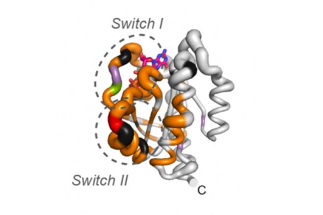
Dyna-1 predicted millisecond-scale movements in regions of KRAS (pictured), a cancer-associated protein that has been notoriously difficult to target. “Switch I” and “Switch II” indicate the protein’s movements in this timescale. The thickness of the circular dashed lines indicates Dyna-1’s predictions compared to the experimentally measured us-ms exchange in color. Credit: Scripps Research
LA JOLLA, CA—Proteins may look like rigid molecular structures, but inside our cells, they are anything but still. They are constantly shifting between different shapes, using these movements to carry out essential jobs like binding molecules and sending signals throughout cells. Some of the most biologically important movements happen in just milliseconds, but predicting them computationally has remained a major challenge.
In a new Nature study published on August 10, 2026, Scripps Research scientists developed Dyna-1, a machine learning (ML) model that can predict protein dynamics on the micro-to-millisecond timescale. In seconds, Dyna-1 identifies regions of proteins likely to undergo these movements, revealing aspects of protein function that have remained hidden from conventional computational approaches. Because these micro-to-millisecond dynamics are particularly conserved across biology since they control function, mapping them could help researchers uncover new connections between protein movement and human health.
“AlphaFold and other language models have completely revolutionized how we predict 3D protein structures, but the key for biology is how fast these proteins move, not just what they look like,” says senior study author Dorothee Kern, Scripps Research professor and a Howard Hughes Medical Institute Investigator. “Proteins that move in the micro-to-millisecond range are the ones most related to biological function, but until now, we didn’t have enough data to simulate these movements on a computer. In this new study, we came up with a really bold idea to get past that.”
Protein dynamics are commonly studied using a technique called nuclear magnetic resonance (NMR), which measures how individual atoms respond to specific stimuli. Unlike techniques that provide a static snapshot of a protein, NMR reveals how proteins move between their different conformations, or shapes.
Kern knew that to achieve what AlphaFold had accomplished for static protein structures, she would need a large amount of experimental NMR data. But there was a major hurdle: this was largely unavailable; they had data for only 133 proteins in the micro-to-millisecond range. Kern and her team would need a much larger dataset—approximately one hundred times more—to develop a viable ML model.
Credit: Scripps Research
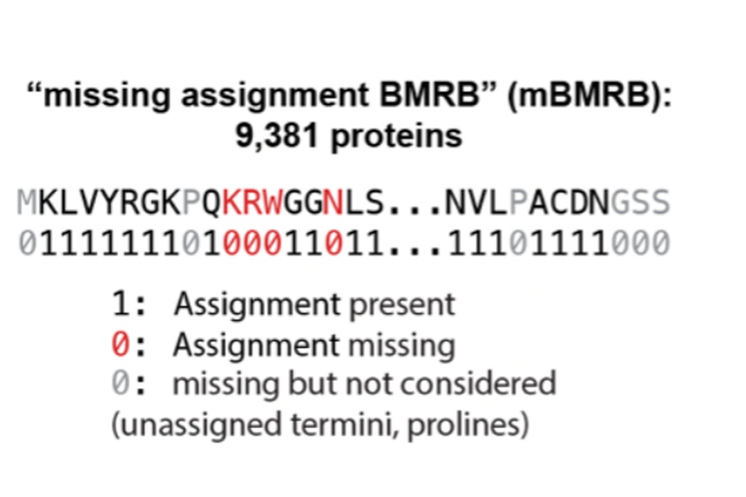
Meanwhile, the Biological Magnetic Resonance Data Bank (BMRB), a public repository of NMR data on biological molecules, contained 10,000 proteins with chemical shift assignments for the majority of the protein, but with some missing assignments, meaning there was no experimental data for certain parts of the proteins. In other words, although these proteins had already been studied using NMR, researchers had been unable to fully interpret the data, leaving potentially useful information hidden in the gaps.
“We were wracking our brains: How do we go from 133 to thousands of proteins? And that’s when we had this bold idea. We would actually use the missing assignments in the BMRB database to our advantage,” Kern adds.
Working with co-first authors Hannah Wayment-Steele and Gina El Nesr, the team proposed that some of these missing assignments could be caused by a phenomenon called “exchange broadening.” When a protein rapidly switches between different conformations, the chemical environment around an atom changes. If those conformational changes occur on the right timescale, the corresponding NMR signal can become so broad that it effectively disappears, meaning a missing signal can itself be evidence that a protein is moving. Of course, not every missing assignment was caused by protein motion (some could have resulted from experimental limitations or other technical issues), but the team built a filter to curate these odd ones out.
This filtering approach left them with roughly 9381 proteins that appeared to have the “exchange-broadened” effect, providing enough data to train an ML model. They set aside the 133 proteins with existing experimental data to test the model’s efficacy later.
They trained several models to predict which parts of a protein would have missing NMR assignments. Dyna-1 performed the best with an accuracy score (or an “AUROC”) of 0.77, meaning it could distinguish protein regions with missing NMR data from those with complete data substantially better than chance (an AUROC of 1 would mean perfect accuracy, while 0.5 would mean the model was no better than guessing). But that was only the first test.
“We were happy with the initial AUROC score, but then the bigger question remained: Has the model learned where proteins move on the millisecond timescale—not just the existence of missing assignments? Has it done transformative learning?” Kern says.
This is where the original 133 proteins came back into the fold. The team tested Dyna-1 against these proteins whose micro-to-millisecond movements had already been experimentally measured. Dyna-1’s performance remained significantly better than the control models, with an overall AUROC of 0.66.
“Notably, correlated motions in the most conserved regions of the protein that are also directly linked to biological function are the best predicted by Dyna-1,” Kern highlights. In one example, the model correctly predicted millisecond-scale movements in regions of KRAS, a cancer-associated protein that has been notoriously difficult to target.
Kern notes that Wayment-Steele and El Nesr developed the model to be straightforward to use, similar to commonly used platforms like AlphaFold and ChatGPT. Researchers across the world are now relying on Dyna-1 to better understand and predict biologically relevant protein dynamics. But Kern and the team are already working on what comes next. Dyna-1 predicts where micro-to-millisecond movement is likely to occur; it doesn’t yet show researchers how exactly a protein moves between its different conformations.
“Dyna-1 predicts for the first time where the motions are on the millisecond timescale, but not how the proteins actually move,” Kern says. “We’re working on Dyna-2 to provide the conformational substrate—the actual protein movements.”
In addition to Kern, Wayment-Steele and El Nesr, authors of the study “Learning millisecond protein dynamics from what is missing in NMR spectra” include Adedolapo Ojoawo of Scripps Research; Ramith Hettiarachchi and Sergey Ovchinnikov of the Massachusetts Institute of Technology; and Hasindu Kariyawasam of Harvard University.
This work was supported by funding from the Howard Hughes Medical Institute (HHMI), the Jane Coffin Childs fellowship and the NSF GRFP.