In a first for cell biology, scientists observe ribosome assembly in real time
November 21, 2019
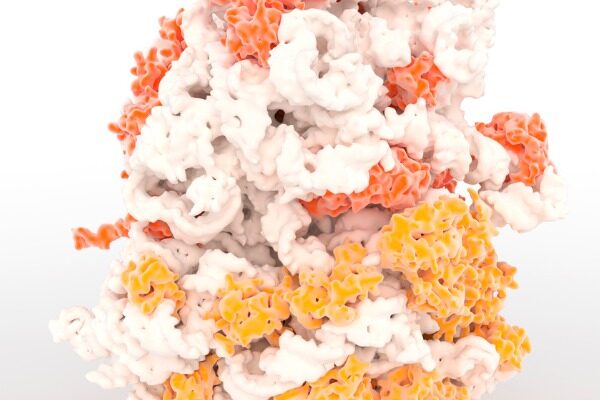
LA JOLLA, CA – A team of scientists from Scripps Research and Stanford University has recorded in real time a key step in the assembly of ribosomes—the complex and evolutionarily ancient “molecular machines” that make proteins in cells and are essential for all life forms.
The achievement, reported in Cell, reveals in unprecedented detail how strands of ribonucleic acid (RNA), cellular molecules that are inherently sticky and prone to misfold, are “chaperoned” by ribosomal proteins into folding properly and forming one of the main components of ribosomes.
The findings overturn the longstanding belief that ribosomes are assembled in a tightly controlled, step-wise process.
“In contrast to what had been the dominant theory in the field, we revealed a far more chaotic process,” says James R. Williamson, PhD, a professor in the Department of Integrative Structural & Computational Biology at Scripps Research. “It’s not a sleek Detroit assembly line—it’s more like a trading pit on Wall Street.”
For the study, Williamson’s lab collaborated with the lab of Joseph Puglisi, PhD, a professor at Stanford University. Although the work is a significant feat of basic cell biology, it should enable important advances in medicine. For example, some current antibiotics work by inhibiting bacterial ribosomes; the new research opens up the possibility of designing future antibiotics that target bacterial ribosomes with greater specificity—and thus, fewer side effects.
More generally, the research offers biologists a powerful new approach to the study of RNA molecules, hundreds of thousands of which are active at any given time in a typical cell.
“This shows that we now can examine in detail how RNAs fold while they are being synthesized and proteins are assembling on them,” says first author Olivier Duss, PhD, a postdoctoral research fellow in the Department of Integrative Structural & Computational Biology at Scripps Research. “This has been a very difficult thing to study in biology because it involves several distinct biological processes that are dependent on each other and have to be detected simultaneously.”
The team used an advanced imaging technology called “zero-mode waveguide single-molecule fluorescence microscopy,” which they have adapted in recent years for real-time tracking of RNAs and proteins. Ribosomes are made of both RNA and proteins, reflecting a molecular partnership that is widely believed to go back nearly to the dawn of life on Earth.
In a proof-of-principle study published last year, the researchers used their approach to record an early, brief and relatively well-studied stage of ribosome assembly from the bacterium E. coli. This involved the transcription, or copying out from its corresponding gene, of a ribosomal RNA, and initial interactions of this RNA strand with a ribosomal protein.
In the new study, the team extended this approach by tracking not only the transcription of a ribosomal RNA but also its real-time folding. The work provided a detailed look at a complex, and until-now mysterious, part of E. coli ribosome assembly—the formation of an entire major component, or domain, of the E. coli ribosome, with assistance from eight protein partners that end up incorporated into the structure.
A key finding was that the ribosomal protein partners guide the folding of the RNA strand through multiple temporary interactions with the strand, well before they nestle into their final places in the folded RNA-protein molecule. The findings, according to the researchers, also hint at the existence of unknown RNA assembly factors, most likely proteins, that were not present in their lab-dish-type imaging experiments but are present in cells and boost the efficiency of RNA folding.
“Our study indicates that in ribosomal RNA-folding, and perhaps more generally in RNA-folding in cells, many proteins help fold RNA though weak, transient and semi-specific interactions with it,” Duss says.
The team will now be able to extend this research further to study not only the rest of ribosome assembly, which involves multiple RNA strands and dozens of proteins, but also the many other types of RNA-folding and RNA-protein interaction in cells.
In principle, this research will yield insights into how RNAs misfold and how such events could be corrected. Scientists believe that many diseases involve or potentially involve the improper folding and related processing of RNAs in cells.
Treatments that already target ribosomes might also be improved. Some current antibiotics, including a class known as aminoglycosides, work by binding to sites on bacterial ribosomes that are not present on human ribosomes. These drugs can have side effects because they also impair the ribosomes of good bacteria, for example in the gut.
“When we understand more fully how bacterial ribosomes assemble and function, we could potentially target them in ways that affect a narrower group of harmful bacterial species and spare the good ones, reducing side effects for patients,” Duss says.
Because ribosomes function as protein makers, they are also crucial to the survival of fast-growing tumor cells. Several classes of cancer drug already work by slowing ribosome formation in one way or another. A better understanding of the human ribosome would, in principle, enable its assembly to be targeted more precisely and potently to block cancer growth, Duss notes.
The other co-author of the study, “Transient Protein-RNA Interactions Guide Nascent Ribosomal RNA Folding,” was Galina Stepanyuk, PhD, of Scripps Research.
Support for the research was provided by the National Institutes of Health (R01 GM051266, R01 GM113078, and R01 GM053757), the Swiss National Science Foundation (P2EZP3-152131, P300PA-160978), and the Human Frontier Science Program (LT000628/2015-L).


Understanding biological factories to fuel drug discovery
Every organism depends on its ability to make cellular proteins from the core set of genetic blueprints—a process carried out in the cell by ancient biological factories. However, when this building process goes awry, various types of disease can take hold. In this opening lecture of the sixth season, Scripps Research professor and regular Front Row host Jamie Williamson unveiled his lab’s unprecedented exploration of these protein-building machines and the secrets they hold in maintaining cellular health. His cutting-edge research is evolving our view of molecular engineering and uncovering new drug targets for bacterial infections and cancer.



Scripps Research neurobiologist Ardem Patapoutian elected to American Academy of Arts and Sciences
Patapoutian, a professor in Scripps Research’s Department of Neuroscience, has catapulted the scientific understanding of sensory transduction—the methods by which the body responds to sensory input from the eyes, tongue, nose and skin.
Chemist Jin-Quan Yu of Scripps Research elected to American Academy of Arts and Sciences
The American Academy of Arts and Sciences, one of the world’s most prestigious honor societies, has named Scripps Research chemist Jin-Quan Yu, PhD, as an elected member of its 2019 class. Yu is known globally for his groundbreaking work in the field of organic synthetic chemistry.
New technique allows study of large molecule assembly in real time
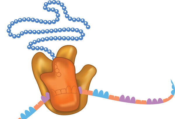
Researchers demonstrate new microscopy technique for studying complex biological processes at the molecular level. The technique allows scientists to observe critical steps in the production of ribosomes, protein-building cellular machines.