Microscopy Core

About the Core
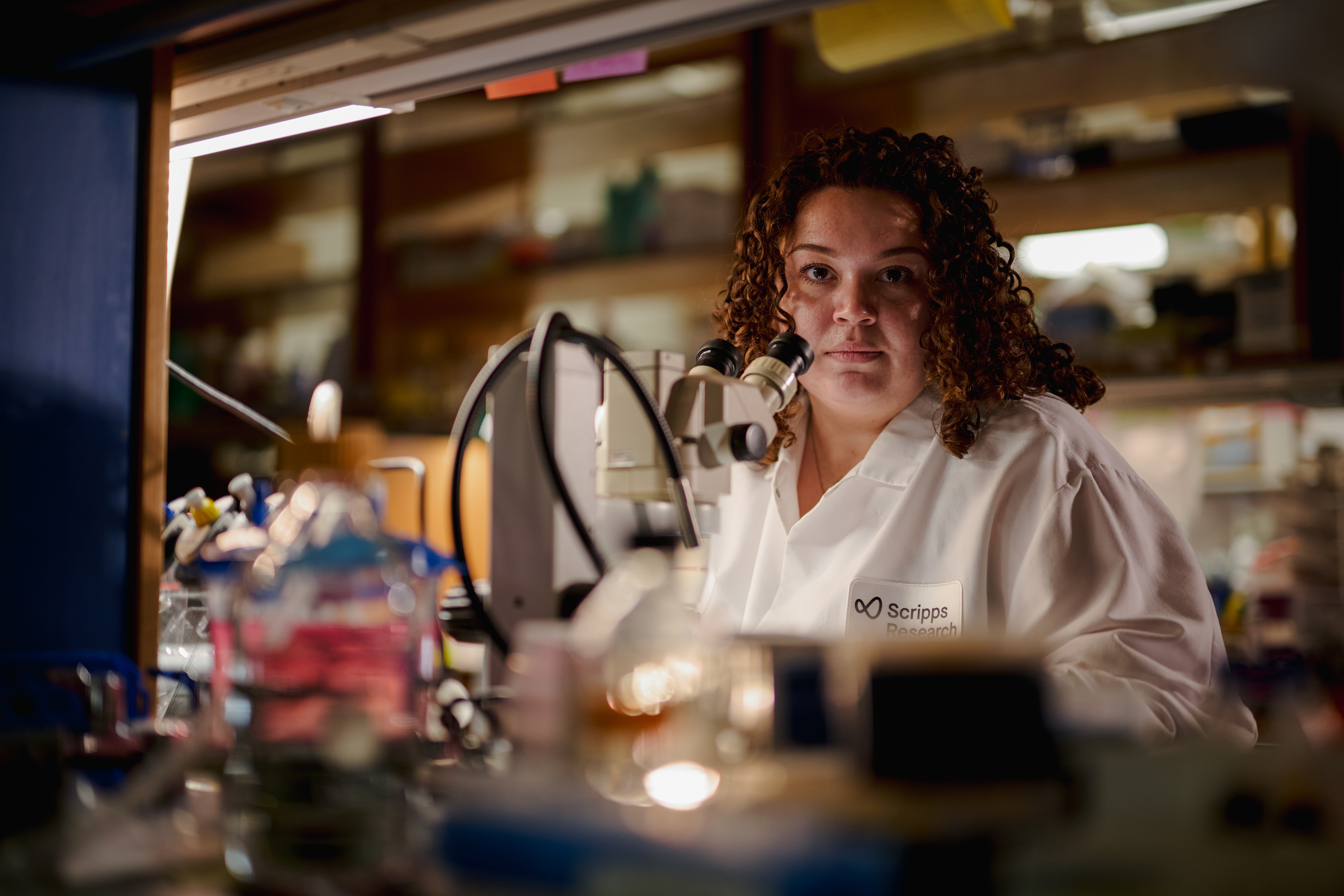
The Microscopy Core facility at Scripps Research provides researchers with equipment and expertise required for multi-dimensional imaging of cells and tissues at high resolution and to perform quantitative image analysis. Resources include widefield, confocal, multi-photon, TIRF, super-resolution (MINFLUX, STED, STORM) and electron microscopy (TEM, SEM), as well as equipment for live cell imaging and image analysis. A cell culture facility (with incubators & biosafety cabinet) is available on site to support live cell imaging. In addition to providing access to advanced instrumentation, this facility, staffed by experts, is a source of instruction and training and constitutes a major educational resource. The Facility staff provides consultation (free for Scripps Research investigators), training, technical support, sample preparation, imaging assistance and collaboration. Services include sample preparation (EM & Histology), molecular localization and image analysis.
Equipment: Electron Microscopy (EM)
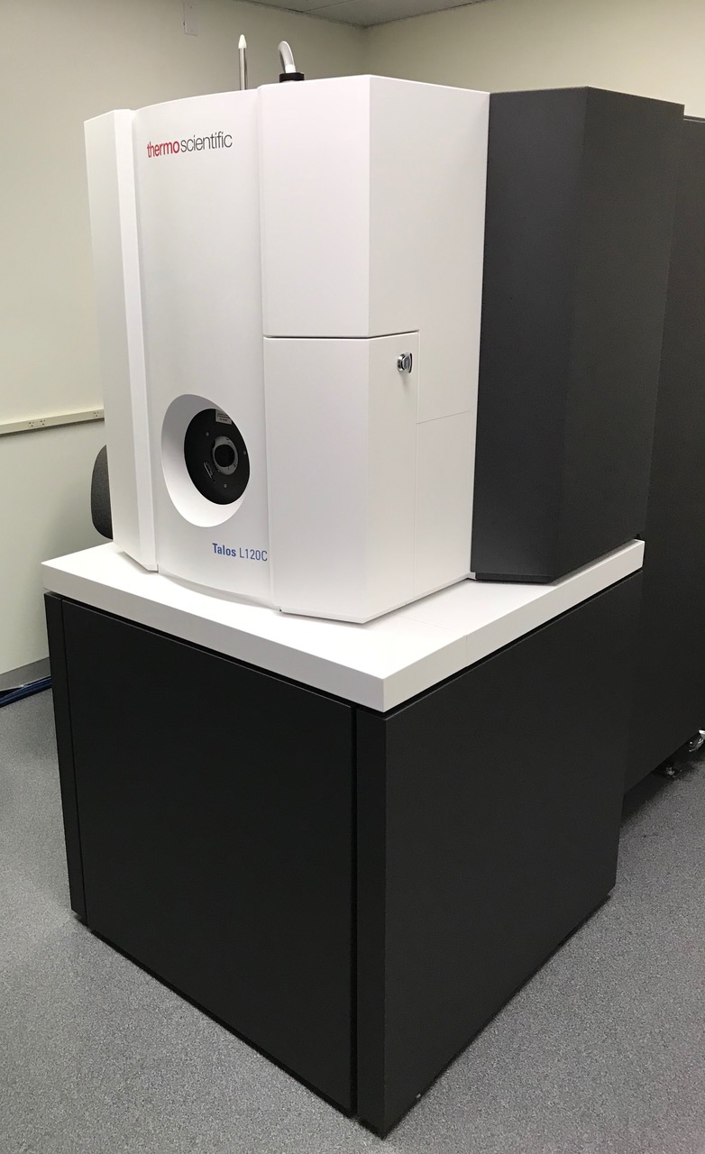
| FEI Talos L120C transmission electron microscope (TEM)
Comes equipped with a C-TWIN lens, cryo box, tomography holder & software and a Ceta 16M CMOS camera. |
 |
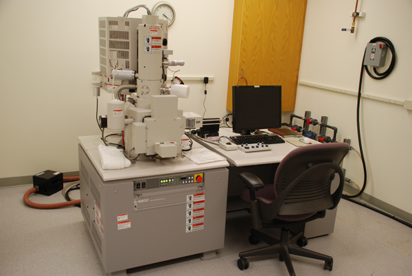
| Hitachi S4800 scanning electron microscope (SEM)
Includes a digital image acquisition, SE detectors, & ExB filter (backscatter capable) and deceleration mode. |
 |
Sample preparation equipment in the EM suite include the following:
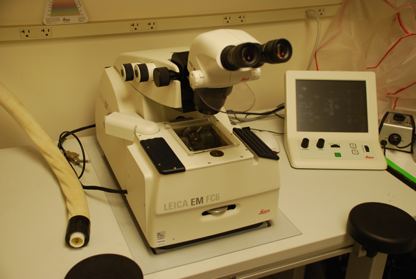
| Leica Ultracut UC6 ultramicrotome
EM FC6 cryochamber |
 |
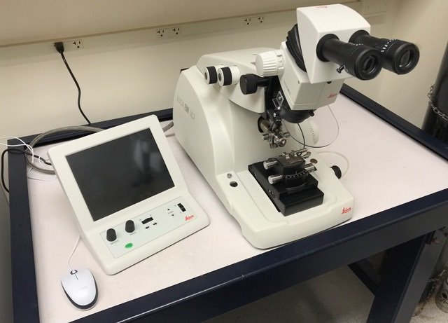
| Leica Ultracut UC7
ultramicrotome
|
 |
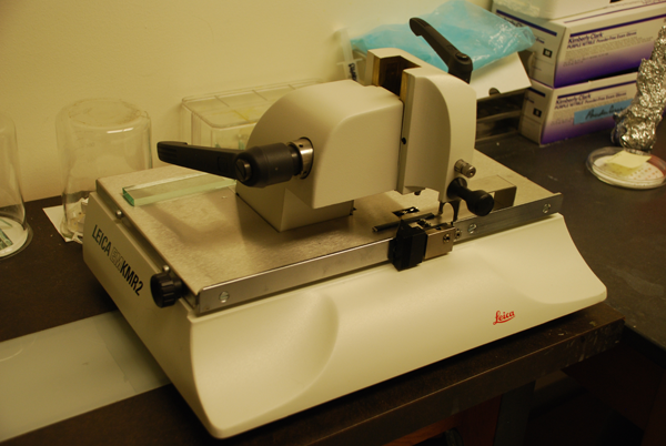
| Leica EM KMR2
glass knife maker |
 |
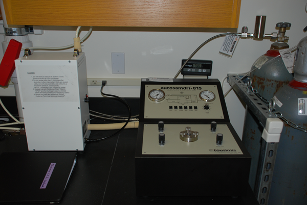
| Tousimis autosamdri 815
critical point dryer |
 |
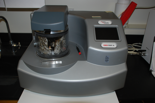
| EMS Q150T S
sputter coater
|
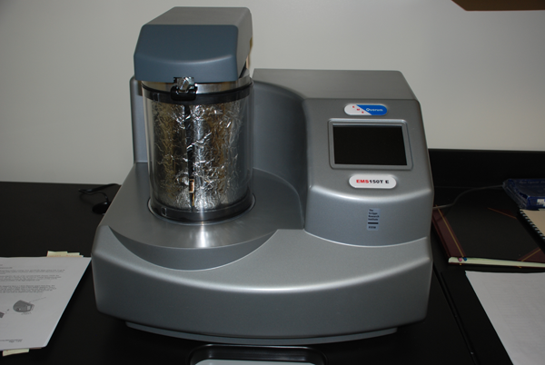 |
| EMS Q150T E
carbon cutter |
 |
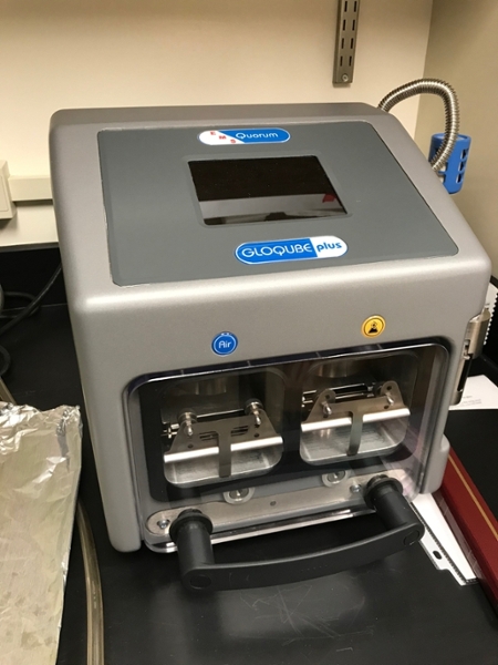
| EMS GloQube Plus
glow discharge unit |
 |
Equipment: Light Microscopy
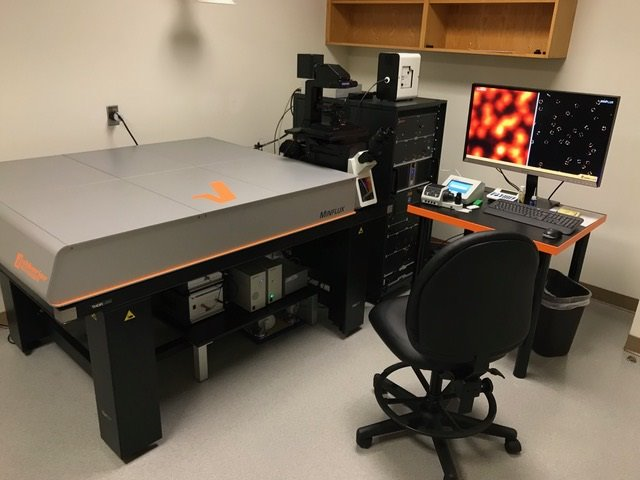
| MINFLUX “Super Resolution” Microscope
|
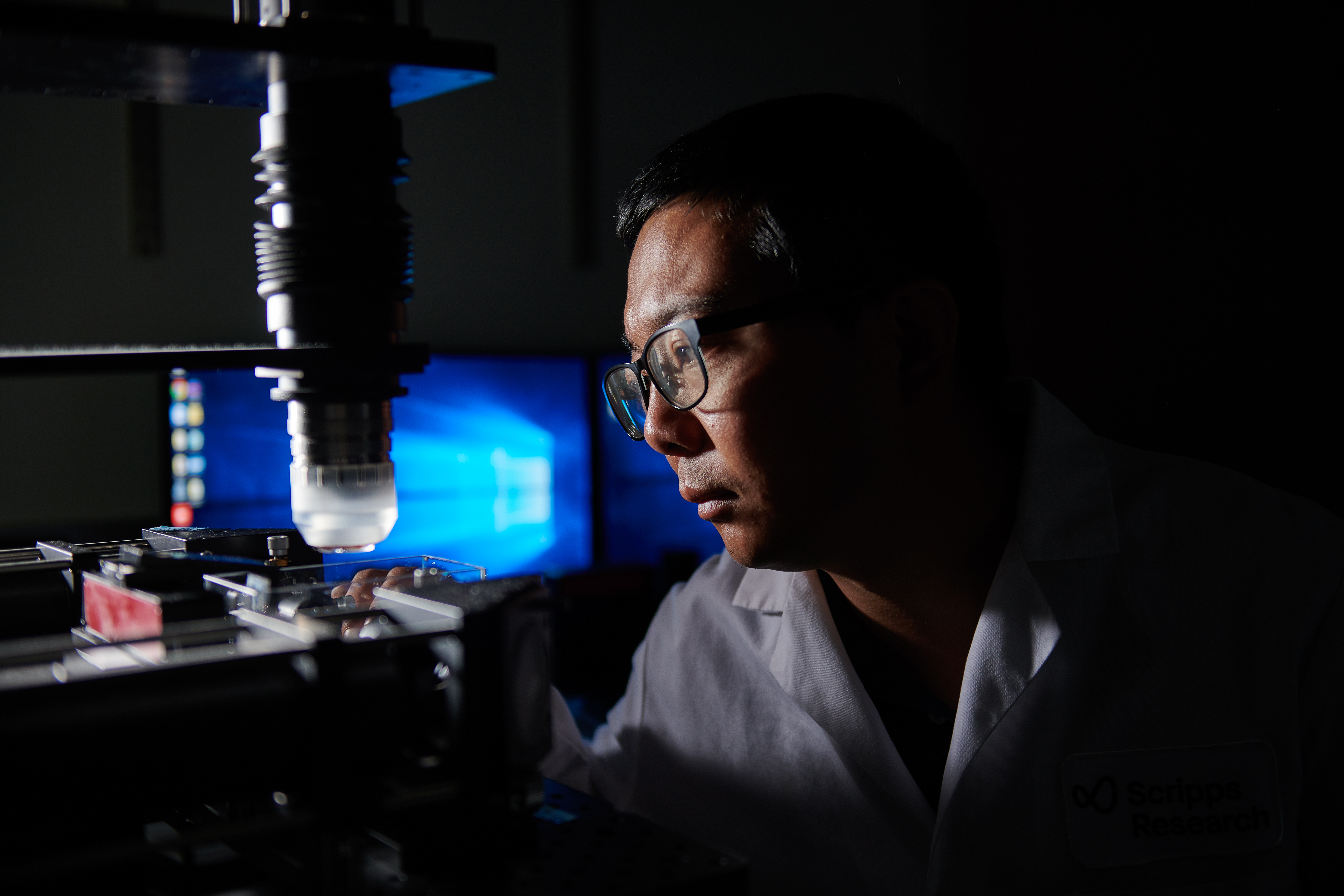
The Scripps Core Microscopy Facility houses an Abberior 3D MINFLUX “super-resolution” microscope. This system, developed in the lab of Dr. Stefan Hell, provides molecular localization precision in fixed samples of approximately 2 nm in all 3 dimensions (X,Y,Z). The system may also be used to track single molecule movements with a temporal resolution as low as 100 µsec. The Scripps Research Core Microscopy Facility’s MINFLUX system was the first one to be installed in North America. The system is equipped with two MINFLUX Lasers (561 nm & 642 nm), adaptive optics, active sub nm accuracy sample stabilization in XYZ, 3D autofocus module, an Olympus IX83 microscope body, four confocal excitation lasers (405, 485, 561, and 640 nm), full auto-alignment, and 5 detection channels. |
| STED “Super Resolution” Microscope | Abberior Facility Line 3D STED/Confocal System equipped with two depletion lasers for STED (775 nm and 595 nm) and five excitation lasers for confocal and STED imaging (405 nm, 440 nm, 485 nm, 561 nm, and 640 nm). Features include Easy3D STED; adaptive illumination (RESCue, DyMIN, MINFIELD); adaptive optics for spherical aberration compensation; RAINBOW spectral detection; MATRIX detector; TimeBow fluorescence lifetime imaging (FLIM); active electronic vibration cancellation; environmental control with stage incubation for live-cell imaging; and Huygens deconvolution software. |
| TIRF / STORM “Super-Resolution” Microscope
|
Nikon TIRF (total internal reflection fluorescence)/STORM (Stochastic Optical Reconstruction Microscopy – “Super-Resolution”) Microscope, equipped with a Hamamatsu Orca Flash 4 CMOS camera, a high power LUNV laser launch with 4 lasers (405/488/561/647 nm), ‘Perfect Focus’ system (laser-based automated focus drift compensation), motorized XY stage, a high resolution piezo-driven Z stage, motorized TIRF optics, fluorescence optics, high NA TIRF objective lenses, DIC optics, and a stage incubator (for live cell imaging).
|
| Multi-photon Laser Scanning Microscopes
|
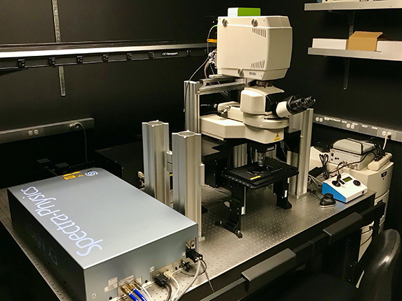
Nikon A1R HD multi-photon laser scanning microscope configured around a large format open frame upright stand to facilitate intravital imaging. The system is equipped with a Spectra-Physics INSIGHT X3DUAL-007 Dual Beam (1300nm tunable / 1040nm fixed) IR Laser (for 2-photon imaging), 4 visible solid state lasers (405 nm, 488 nm, 561 nm, and 640 nm) for confocal imaging, 4 non-descanned detectors (3 GaAsP and 1 high sensitivity PMT), 4 descanned detectors (2 GaAsP and 2 high sensitivity PMTs), AOTF, motorized XYZ translation stage, nosepiece objective piezo for fast Z-stacking, and multiple high NA LWD dipping objective lenses (10x, 16x, 25x, 60x). |
| Laser Scanning Confocal Microscopes
|
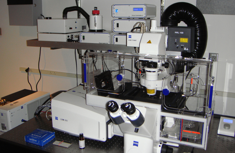
Zeiss LSM 880 Airyscan confocal laser scanning microscope configured around an inverted stand and equipped with a 32-channel array detector (Quasar) plus 2 side PMT detectors, 32 GaAsP PMT array (Airyscan detector for enhanced resolution), a transmitted light detector, AOTF, ‘Definite Focus’ system (laser-based automated focus drift compensation), a motorized XY stage, piezo driven Z stage, a PeCon full stage incubator (with regulation for temperature, humidity, CO2), multiple high NA objective lenses (10x, 20x, 40x oil & 63x oil, 100x oil), DIC, and 7 laser lines: 405, 458, 488, 514, 561, 594 and 633 nm. The system is set up for FRET and FRAP analysis. |
| Laser Scanning Confocal Microscopes
|
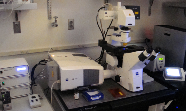
Zeiss LSM 780 confocal laser scanning microscope configured around an inverted stand and equipped with a 32-channel GaAsP array detector (Quasar) plus 2 side PMT detectors, a transmitted light detector, ‘Definite Focus’ system (laser-based automated focus drift compensation), AOTF, a Märzhäuser motorized XY stage, a Pathology Devices stage incubator (with regulation for temperature, humidity, CO2), multiple objective lenses (10x, 20x, 40x oil, 40x water, 63x oil, 63x water, and 100x oil), DIC, and 7 laser lines (405nm, 458nm, 488nm, 514nm, 561nm, 594nm, 633nm). The system is set up for FRET and FRAP analysis. |
| Laser Scanning Confocal Microscopes
|
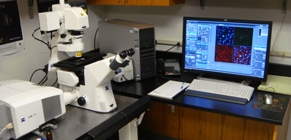
Zeiss LSM 710 confocal laser scanning microscope configured around an inverted stand and equipped with a 3 PMT detectors, external “BiG” (binary GaAsP) detectors, a transmitted light detector, ‘Definite Focus’ system (laser-based automated focus drift compensation), AOTF, a Märzhäuser motorized XY stage, a Pathology Devices stage incubator (with regulation for temperature, humidity, CO2), multiple objective lenses (5x, 10x, 20x, 40x oil, 40x water, 63x oil & 100x oil), DIC, and 7 laser lines: 405, 458, 488, 514, 561, 594 and 633 nm. The system is set up for FRET and FRAP analysis. |
| Widefield Fluorescence Microscopes
|
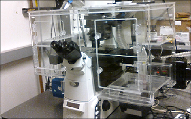
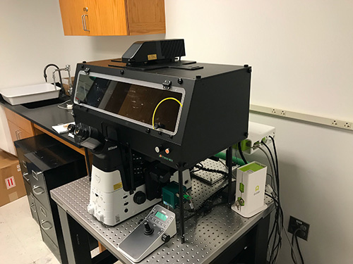
Nikon Ti2-E Inverted Motorized Microscope equipped with Ti2-ND-P Perfect Focus System 4, TI2-S-SE-E Motorized Stage with Encoder, 5 objective lenses (10x, 20x, 40x water, 60x oil, 100x oil), DIC optics, fluorescence (blue, green, red, far red) optics, Photometrics IRIS 15 sCMOS Camera, Tokai Hit Incubation System, and multiple stage adaptors (for slides, dishes, and multiwall plates). Attached to this microscope is an Alvéole PRIMO micropatterning system for generating customized cellular microenvironments. |
| Widefield Fluorescence Microscopes
|
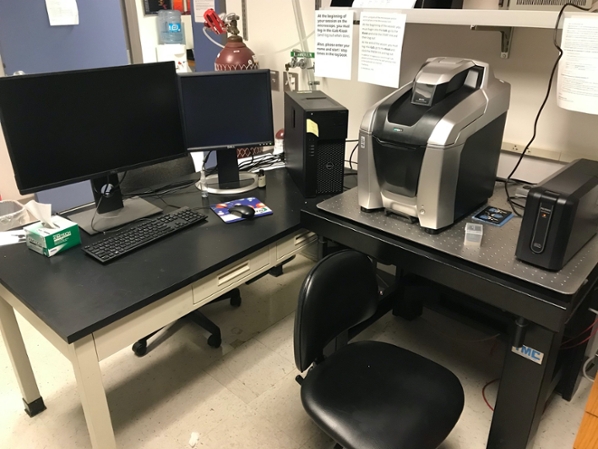
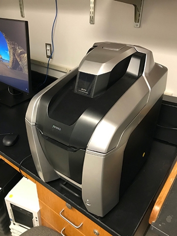
Keyence BZX710 all-in-one fluorescence microscope equipped with a cooled CCD camera 2x, 10x, 20x, 40x, 60x oil, and 100x oil objective lenses, brightfield and fluorescence (blue, green, red, far red) optics, auto-focus, a motorized stage (X, Y, and Z), a live cell incubation module (stage incubator) and multiple stage adaptors (for slides, dishes, and multiwall plates). |
| Widefield Fluorescence Microscopes
|
Keyence BZX710 all-in-one fluorescence microscope equipped with a 4K cooled CCD camera 2x, 10x, 20x, 20x lwd, 40x, and 60x oil objective lenses, brightfield, phase, and fluorescence (blue, green, red, far red) optics, auto-focus, a motorized stage (X, Y, and Z), and multiple stage adaptors (for slides, dishes, and multiwall plates). |
| Spinning Disc Confocal Microscope
|
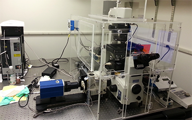
Nikon spinning disc confocal microscope equipped with a Yokogawa CSU-21 spinning disc, upgraded with a Spectral Applied Research Borealis system with a 4-line laser launch (405, 488, 561, and 642 nm), a CoolSnap HQ2 camera, 5 objective lenses (10x, 20x, 40x oil, 60x oil, 100x oil), DIC, phase contrast, and a stage incubation system (to support live cell imaging). |
| Slide Scanners
|
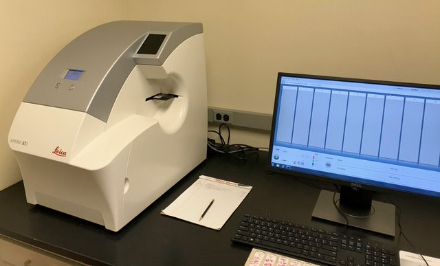
Leica AT2 whole slide scanner (brightfield) with 400 slide capacity, 20x/0.75 NA Plan Apo objective lens (and a 2x optical magnification changer for 40x scanning). (pictured)
Vectra Polaris Automated Quantitative Pathology Imaging System, for fluorescence and brightfield whole slide scanning, equipped with Plan-Apo objective lenses (10x/0.20 NA, 20x/0.45 NA, 40x/0.75 NA), XYZ motorized stage, fluorescence and transmitted light optics, multispectral imaging (440 – 720 nm), 5 filter cubes (FITC, Cy3, DAPI, Texas Red, ET-Cy5), 80-slide capacity, and inForm Tissue Finder software (for automatic detection, segmentation, and analysis of slides). |
| Image Analysis
|
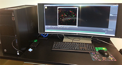
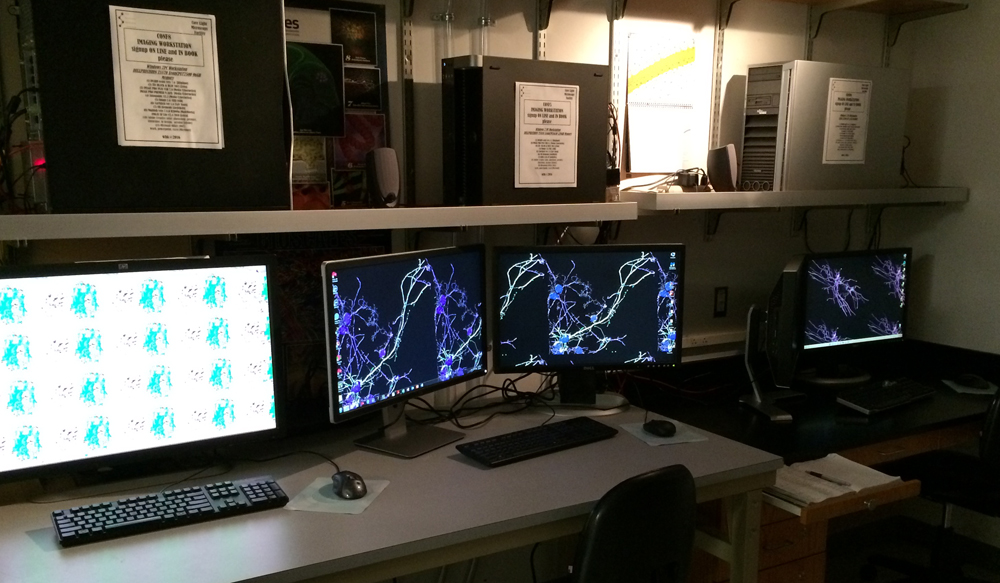
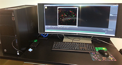
Multiple computer workstations with a variety of image analysis programs, including: Huygens, Image Pro Premier, ImageJ, IMARIS, LAS AF Lite, NIS-Elements, Zen (Black & Blue).
|