


- VOL 18. ISSUE 5
March 02, 2018
SEARCH NEWS & VIEWS

Cross-bred flies reveal new clues about how proteins are regulated
'Click chemistry' reactions may boost cancer-fighting drug potency
Decoding the genome’s dark matter
TSRI scientists receive $15 million to study viral outbreak survivors
Next-generation arthritis treatments could benefit both horses and humans
NEWS & VIEWS HOME
PAST ISSUES
KUDOS
SCIENTIFIC CALENDAR
CA AUDITORIUM EVENTS
CONTACT

- The San Diego Union-Tribune
Scripps Research stroke drug shows effectiveness, safety - GenomeWeb
Researchers Build Map of Sequence Constraints for Human Genome


FOLLOW US


Of Note
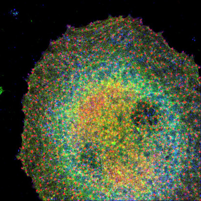
TSRI scientists win international photo contest
Three biologists at The Scripps Research Institute (TSRI) have been honored for their skill in capturing eye-catching images in the lab. Researchers Roberta Nowak, Kasturi Pal, PhD, and Catherine Cheng, PhD, have been named winners in the Vector Laboratories 2017 Photo Contest.
All three winners are members of Velia Fowler’s, PhD, Laboratory for Cellular Architecture, and their images show the amazing range of research at TSRI.
This image by Nowak and Pal shows mouse megakaryocyte spreading on collagen coated cover slip. Single optical section of Airyscan stack showing myosin-GFP (green) signal along with phalloidin (red) and vinculin (blue).
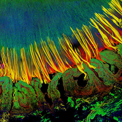
The lens is a transparent and avascular organ in the front of the eye that is responsible for focusing light onto the retina in order to transmit a clear image. In humans, ciliary muscles contract to change the tension of zonule fibers to deform the lens, leading to an increase in the lens optical power to focus on nearby objects, a process known as accommodation. This image from Cheng shows a 3D reconstruction of a confocal z-stack through a human lens with attached cilliary body and zonules. The zonules are stained with wheat germ agglutinin (red) and microfibril-associated glycoprotein MAGP-1 (yellow). The ciliary body (foreground) and the lens (behind the zonules) are stained with phalloidin (F-actin, green) and DAPI (nuclei, blue).
Send comments to: press[at]scripps.edu
- CONNECT
- VISIT
- CA Campus:
- • Maps & Directions
- • Parking
- FL Campus:
- • Maps & Directions
- • Parking
- RESEARCH
- WORK
- CHALLENGE
California: 10550 North Torrey Pines Road, La Jolla, CA 92037 - (858) 784-1000
Florida: 130 Scripps Way, Jupiter, FL 33458 - (561) 228-2000
Copyright © 2024, The Scripps Research Institute (TSRI). All Rights Reserved.