


- VOL 15. ISSUE 16
May 11, 2015
SEARCH NEWS & VIEWS

Study Finds Metabolic Link Between Bacterial ‘Biofilms’ and Colon Cancer
Researchers Connect Haywire Protein to Breast Cancer, Leukemia
Married to Science
Scientists Win $2.4 Million to Expand Development of New Pain Therapies
NEWS & VIEWS HOME
PAST ISSUES
KUDOS
SCIENTIFIC CALENDAR
CA AUDITORIUM EVENTS
CONTACT

- Laboratory Equipment
Scientists Discover Clues to Altered Brain Wiring in Autism - The San Diego Union-Tribune
Our perpetually vigilant internal guardian


FOLLOW US


Study Finds Metabolic Link Between Bacterial ‘Biofilms’ and Colon Cancer
A team led by scientists at The Scripps Research Institute (TSRI) and Johns Hopkins University School of Medicine has uncovered a big clue to how bacteria may promote some colon cancers.
The study, reported in Cell Metabolism on May 7, 2015, used novel metabolomic technologies to reveal molecular evidence suggesting a vicious circle in which cancerous changes in colon cells promote the growth of bacterial conglomerations called biofilms, and biofilms in turn promote cancer development.
On the whole, the findings suggest that removing bacterial biofilms could be a key strategy for preventing and treating colon cancers, which currently kill about 50,000 Americans per year. The study also revealed an apparent metabolic marker of biofilm-associated colon cancers.
A Wide Net
The research, which used sophisticated “metabolomics” techniques, was a collaboration between groups led by Gary Siuzdak, professor of chemistry, molecular and computational biology and senior director of the Scripps Center for Metabolomics at TSRI, Cynthia L. Sears, professor of medicine, oncology and molecular microbiology and immunology at the Johns Hopkins University School of Medicine and Bloomberg School of Public Health, and David Edler, associate professor at the Karolinska Institute.
A previous study led by Sears and colleagues provided evidence that the tissue in and around cancers of the ascending colon, on the right side of the abdomen, almost always harbors bacterial conglomerations called biofilms.
“In the current study, we wanted to understand more about what was happening,” said Caroline H. Johnson, member of the Scripps Center for Metabolomics and co-first author of the new report with Christine M. Dejea of Johns Hopkins. “In particular, we wanted to determine if there was a metabolic link between the biofilm and colon cancer.”
Metabolites are small molecules in blood and tissues that are products of the myriad metabolic processes in cells. More than 10,000 distinct metabolites normally can be found in humans.
The team began the search with an “unbiased screen,” a wide-net technique—using advanced liquid chromatography and mass spectrometry and their XCMS metabolomic cloud-based platform—that registered the levels of thousands of metabolites in a set of colon tissue samples from patients at Johns Hopkins and at the Karolinska Institute in Sweden.
The data showed that polyamines were important in general and one metabolite—N1, N12-diacetylspermine—was particularly prominent, on average about nine times more abundant in cancerous tissue, compared to nearby non-cancerous tissue.
A Vicious Circle
In further tests, the team found that even among cancerous samples, the same metabolite was four times more abundant in the presence of biofilms. In other words, the cancerous cells and the biofilms both seemed to be contributing to its overproduction.
With a sophisticated technique called “nanostructure imaging mass spectrometry” (NIMS), the team was able to map the precise locations of N1, N12-diacetylspermine in tissue samples, confirming its higher levels in both tumors and biofilms.
The researchers also carried out a technique called “global isotope metabolomics,” using an isotope of N1, N12-diacetylspermine to trace its metabolic fate in cells in an unbiased manner, finding that it appears to be a metabolic end-product.
That colon tumors would produce abnormally high amounts of N1, N12-diacetylspermine is not surprising. The molecule belongs to a family of metabolites called polyamines, which are known to have roles in driving cell growth and which are commonly upregulated in cancers as well as in healthy fast-growing tissues. N1, N12-diacetylspermine itself has been observed at higher levels in colon cancer and is considered a potential biomarker for early cancer diagnosis.
But why would bacterial biofilms also be linked to higher levels of N1, N12-diacetylspermine? It turns out that bacteria, too, use polyamines to drive their own cells’ proliferation and to build biofilms. Polyamines are such ancient, ubiquitous molecules that bacteria apparently can even use those produced by their animal hosts.
Thus, biofilms may promote cancer in the colon by inducing chronic inflammation and associated cell proliferation. That increased cell proliferation would be accompanied by a rise in the production of polyamines. Resident bacteria, in turn, could use this abundance of polyamines to make more biofilms—completing the vicious circle. Along the way, levels of the by-product N1, N12-diacetylspermine would be driven higher and higher.
The Way Forward
The researchers now want to find out more about the molecular pathways through which polyamines contribute to tumor growth and biofilm construction. They want to know as well why bacterial biofilms are found frequently in association with tumors of the ascending colon, but less frequently in tumors further along in the colon.
“We’d also like to look at samples from other populations that have a low level of colon cancer and different traditional diets, because we know that diet can influence polyamine levels,” said Johnson.
In the meantime, treatment with antibiotics may be an option for removing colonic biofilms and reducing the cancer risks they bring. The scientists found that colon cancer samples from patients who had taken oral antibiotics 24 hours prior to surgery harbored no biofilms and no cultivable bacteria and exhibited significantly less N1, N12-diacetylspermine, on average, than samples from patients who had not taken antibiotics.
In addition to Siuzdak, Sears, Johnson and Dejea, other co-authors of the paper, “Metabolism links bacterial biofilms and colon carcinogenesis,” were Linh T. Hoang, Antonio F. Santidrian, Brunhilde H. Felding and Winnie Uritboonthai of TSRI; Laura Goetz of University of California, San Diego; Elizabeth C. Wick, Elizabeth M. Hechenbleikner, Robert A. Casero Jr. and Drew M. Pardoll of Johns Hopkins; David Edler of Karolinska University Hospital; Gary J. Patti and Kevin Cho of Washington University School of Medicine in St. Louis; and James R. White of Resphera Biosciences. For more information, see http://www.cell.com/cell-metabolism/home
Support for the research came in part from the California Institute of Regenerative Medicine (TR1-01219); the National Institutes of Health (R01 CA170737, R24 EY017540, P30 MH062261, RC1 HL101034, P01 DA026146, R01 CA151393, R21 CA170492, K087856, P30 DK089502, P30 CA006973, R01 CA051085, R01 CA098454 and T32AI007417) and the U.S. Department of Energy (FG0207ER64325 and DE-AC0205CH11231).
Send comments to: press[at]scripps.edu
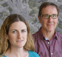
Authors of the new study included Caroline Johnson and Gary Siuzdak of TSRI's Center for Metabolomics. (Photo by Cindy Brauer.)