


- VOL 14. ISSUE 19
June 30, 2014
SEARCH NEWS & VIEWS

Letter from Dick Gephardt, Chair of the TSRI Board of Trustees
Team Finds Potential New Use for Cancer Drug in Gene Therapy for Blood Disorders
Scientists Pinpoint How Genetic Mutation Causes Early Brain Damage
Study Finds Molecular ‘Yin-Yang’ Regulates Blood Vessel Growth
In Memoriam: Carlos F. Barbas III (1964 – 2014)
Scientific Art on the Fly: Behind the Scenes of the New Airport Exhibit
NEWS & VIEWS HOME
PAST ISSUES
KUDOS
SCIENTIFIC CALENDAR
CA AUDITORIUM EVENTS
CONTACT

- Laboratory Equipment
Scientists Discover Clues to Altered Brain Wiring in Autism - The San Diego Union-Tribune
Our perpetually vigilant internal guardian


FOLLOW US


Scientific Art on the Fly: Behind the Scenes of the New Airport Exhibit
By Madeline McCurry-Schmidt
The Scripps Research Institute (TSRI) is sharing biology with a brand new audience. Starting this month, visitors arriving at the San Diego International Airport will be greeted by an exhibit titled "Taking Art to the Cellular Level," in the new part of Terminal 2.
Among the pieces on display are a number of images created by TSRI scientists, including Lab Assistant Adam Gardner and Senior Research Associate Ludovic Autin of Professor Arthur Olson’s lab. Here, Gardner and Autin share their thoughts on their pieces, the exhibit and the role of visual art in the sciences.
News&Views: What did you depict in your images, “Our Cellular Genesis (Nucleus and ER)” and “Mycoplasma”?
Gardner: “Our Cellular Genesis” is a kind of fantasy scape. You see the nucleus and you see the ER [endoplasmic reticulum], and three-fourths of the ER is cut away, so it almost looks like a cloud if you’re blurring your eyes—like a cloud and a sun. It was a piece of a larger illustration of a full cell with lipid membrane proteins, but we took that piece out and put a grey background behind it.
Autin: For the other piece, our colleague David Goodsell [Associate Professor of Molecular Biology at TSRI] made a painting of a mycoplasma, which is one of the smallest organisms you can find. It’s an interesting organism because it is the first that has been completely synthesized—meaning that we know its genome and we can synthesize it completely. So we have a lot of information—what protein is inside, how it works. The interesting thing is that there’s no nucleus, so the DNA is not compact. We were looking at this painting and said, “Okay, let’s try to do it in three dimensions.”
N&V: How do you choose which colors to use?
Gardner: I usually pick certain colors and then different lightness and darkness values of those colors for the different elements. That way, you have the same color scheme, but can tell pieces apart. If there’s more than one color in the scene, I do the same thing. I’ll have a blue that I like and a red that I like and then I’ll use different shades of each color.
N&V: What are your backgrounds?
Autin: I have a PhD in molecular modeling.
Gardner: I’m self-taught in 3D. When I was in high school, I would try to become like a guy at Pixar, making movies. When I went to college, I thought, “This is not a good way to make a living.” I love biology and chemistry, so I got a bachelor’s degree in those subjects, graduated, found out about Professor Art Olson’s molecular graphics lab, saw that I could merge the two disciplines that I love—and here I am!
N&V: What tools make this work possible?
Autin: There is a huge amount of software from the game and movie industries. I try to merge them with molecular modeling software, taking what is best in both fields—to use the same software but for a different purpose.
Gardner: Ever since the birth of the field of computer graphics, there has been a lot of collaboration between scientists and the game and movie industries. There has always been a weird cross-over.
N&V: What do you hope people notice when they walk past your piece in the airport?
Gardner: Most images in the exhibit came from microscopy, so they are real images. Our images were the only ones to involve computation. There are merits to computation, so it’s nice to see that represented.
Autin: It’s also nice to give people an idea of how cells look. I think that in some people’s minds, the cell is just a sphere, but it’s actually atoms everywhere.
N&V: How do you think these sorts of models help scientists?
Autin: These computer generated models synthetize different imaging data and provide a picture of the crowded environment of the cell, which no current experiment can produce. These models can help scientists evaluate and define new hypotheses.
Gardner: Like what Autin said, these models provides a view of the cell in a way that you can't see using just one source of traditional imaging data.
N&V: How could these images be useful for the public?
Gardner: A pretty picture draws your attention, and then prompts questions. It’s like an advertisement for science. If it looks appealing enough, hopefully some kid somewhere will say, “That looks cool, I want to be a scientist!” or “That looks cool, I want to understand more.”
N&V: What projects do you have coming up?
Autin: Art Olson recently got a grant to find out if 3-D tangible models associate with augmented reality [AR] could help high school students better understand structural biology. So we are working to see if it can.
Gardner: Autin is a really wonderful coder, and he’s doing all the AR parts of it and using game engines, like Unity, to import it into iOS.
Send comments to: press[at]scripps.edu
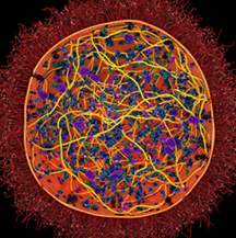
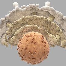
“Mycoplasma” (top) by Lab Assistant Adam Gardner and Research Associate Ludovic Autin and “Our Cellular Genesis (Nucleus and ER)” by Gardner are part of an exhibit in the San Diego Airport titled "Taking Art to the Cellular Level."