TSRI method accelerates studies on carbohydrate biology
Nearly every living cell is studded with branching chains of carbohydrates called glycans. Glycans play diverse roles in shaping how a cell interacts with its environment.
Now, researchers at The Scripps Research Institute (TSRI) have described a new method for adorning cells with various glycans and screening interactions between glycans and proteins. Their breakthrough, published recently in Nature Communications, may expand research on the roles of glycans in human diseases, including cancers.
“Scientists have been trying to make glycan arrays that every scientist interested in glycans can access in their own labs for years,” says Peng Wu, PhD, a TSRI associate professor and senior author of the study. “We’ve not only done it, but we’ve done it in a way that’s very easy.”
Researchers solve problem in glycan screening
The patterns of glycans and glycan-binding proteins on a cell’s membrane can differentiate cancer cells from healthy cells, control cells’ roles in development and contribute to diverse interactions between adult cells. Genetic diseases that affect the ability of cells to properly create glycans can shorten lifespan and lead to musculoskeletal problems.
But studying glycans has been notoriously tricky. While scientists know how to synthesize diverse proteins and DNA molecules in the lab, creating glycans on demand has been chemically challenging.
To study which proteins in a cell interact with glycan molecules, researchers have typically turned to glycan binding arrays, in which dozens or hundreds of glycans are attached to a glass slide. Researchers then expose the slide to cells or proteins of interest and observe whether the cells or proteins stick to the glycans on the slide. But making these arrays is time-consuming and expensive.
“In the past, if you wanted to make an array with 100 sugars, then you had to chemically synthesize 100 sugars individually, which can be difficult,” says Wu. “Only specialized carbohydrate chemists can make them in certain labs.”
Wu and his colleagues instead decided to harness the power of the enzymes cells use naturally to produce glycans. These enzymes work in a stepwise fashion to create branching glycans— one small piece of a sugar is made by one specialized enzyme, then another enzyme creates the next branch in the chain, and so on. The researchers found that even structurally-related unnatural sugars can be added in this way.
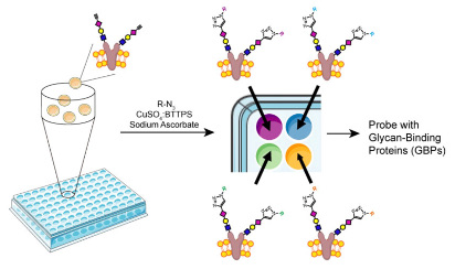
Wu’s team began with mutated rodent ovary cells that had a very narrow repertoire of glycans on their surface. This was a simpler system than using human cells with many types of glycans. The researchers then exposed the cells to different sets of glycan-creating enzymes to control the addition of carbohydrate branches to the glycans on each cell.
With this method, they created cell arrays each studded with different glycans, including unnatural ones.
“The only limitation is the enzymes that we have available, and the fact that you have to start with cells that already have simple glycosylation,” says Wu. “But we were able to create all the glycans we wanted.”

(Image from Wu Lab)
Putting the library to the test
To test the utility of the new cell array, Wu and his colleagues screened an array of cells, each displaying a different glycans, to determine which ones bound to Siglec-15, a known glycan-binding protein that plays a role in bone development and remodeling. Siglec-15 is considered a potential target for drugs treating postmenopausal osteoporosis, so understanding how it interacts with carbohydrates is critical. The team identified three structures with strong binding to Siglec-15.
The researchers then incubated human osteoprogenitor cells with mutated rodent ovary cells displaying one of the three structures during differentiation. The team found that this process suppressed the formation of osteoclasts, a Siglec-15-expressing bone cell that absorbs bone tissue during growth and healing. This finding reinforces the idea that Siglec-15 is a good target for osteoporosis treatments, and that the new glycan screening strategy can point researchers to promising new drugs.
“We don’t know if this will be used in the broad community—it depends on the availability of enzymes and cells,” says Wu. “But if a whole bunch of cells with simple and homogeneous glycans can be made available, that would be huge for the field.”
In addition to Wu, authors of the study, “Cell-Based Glycan Arrays for Probing Glycan-Glycan Binding Protein Interactions,” include Jennie Briard and Matthew Scott Macauley of The Scripps Research Institute; Hao Jiang of the Ocean University of China; and Kelley Moremen of the University of Georgia.
This work was supported by funding from the National Institutes of Health (GM093282, GM113046 and GM103390) and the NSFC-Shandong Joint Fund for Marine Science Research Centers (U1606403).
Send comments to: press[at]scripps.edu


