


- VOL 15. ISSUE 11
March 30, 2015
SEARCH NEWS & VIEWS

Cells You Can See: A Profile of Art Olson
Team Breaks Imaging Barrier
Enzyme Keeps Blood Stem Cells Functional to Prevent Anemia
NEWS & VIEWS HOME
PAST ISSUES
KUDOS
SCIENTIFIC CALENDAR
CA AUDITORIUM EVENTS
CONTACT

- Laboratory Equipment
Scientists Discover Clues to Altered Brain Wiring in Autism - The San Diego Union-Tribune
Our perpetually vigilant internal guardian


FOLLOW US


Team Breaks Imaging Barrier
Advances in Electron Microscopy Could Aid Drug Design
By Madeline McCurry-Schmidt
Scientists at The Scripps Research Institute (TSRI) have broken a major barrier in structural imaging. Their study, published recently in the journal eLife, shows a protein complex at the highest resolution ever achieved with a standard technique called single particle cryo-electron microscopy.
“The instruments and software are now so good that we do not know what the barriers are any more,” said Bridget Carragher, a professor at TSRI with a joint appointment at the New York Structural Biology Center.
With single particle cryo-electron microscopy, scientists freeze a sample and then expose it to a beam of high-energy electrons. This excites electrons in the sample, allowing scientists to capture an image.
While the technique has many practical advantages over other structural biology methods, scientists have so far not been able to reach resolutions more detailed than 3 Angstroms (one ten-billionth of a meter, marked with the symbol Å). At this resolution, some of the details of the structure that are important for guiding drug design are not discernable.
The new study shows that reaching resolutions greater than 3 Å is possible using single particle cryo-electron microscopy. The imaged protein complex reveals individual molecules at 2.8 Å and is, to the researchers’ knowledge, the first time a paper has been published showing individual water molecules using this technique.
Better Imaging, Better Drugs
The scientists used a new type of electron microscope, called the FEI Titan Krios, and a new-generation camera, called a Gatan K2 Summit, to break the 3 Å barrier.
The FEI Titan Krios is housed on TSRI’s La Jolla, California, campus. It has a higher energy electron source and a more stable platform than other types of electron microscopes. It also operates with software developed at TSRI through the National Resource for Automated Molecular Microscopy to find the best parts of a sample for imaging.
The Gatan K2 Summit camera improves imaging by directly detecting electrons, instead of losing resolution by converting electrons to light. The camera can also capture a series of images, essentially a movie, giving scientists the ability to correct for movements in the specimen and make the images as sharp as possible.
Revealing high-resolution details in a structure helps researchers develop new drugs to treat disease. Structures seen at greater than 3 Å might show vulnerabilities in a virus where drugs could bind, for example.
“By seeing everything in more detail, you can design more effective drugs,” said Melody Campbell, a TSRI graduate student and co-first author of the new paper with David Veesler, previously a post-doctoral fellow at TSRI and now an assistant professor at the University of Washington.
The advances in single particle cryo-electron microscopy also allow scientists to image more kinds of structures, more quickly. For many years, scientists have relied on a high-resolution imaging technique called X-ray crystallography. Although X-ray crystallography has led to many advances in drug design, figuring out how to grow a crystal can take years and not all structures can be crystallized.
Electron microscopy does not require a crystal, however, and many projects take only weeks or months.
In the new study, the researchers imaged a protein complex from a microbe called Thermoplasma acidophilum. This protein complex, called a proteasome, is also found in humans and is an important target for treating many types of cancer.
The team spent several months setting up the instruments—since the FEI Titan Krios was new to the institute—and then they captured all the raw data over a single weekend. They then used computational programs to select the clearest images and refine them over several months to build a 3D model of the proteasome.
“It was a relief to know we had finally done it,” said Campbell. “Now we hope other people can just hop on the microscope, use similar strategies and also get high-resolution structures.”
In addition to Carragher, Campbell and Veesler, authors of the study, “2.8 Å resolution reconstruction of the Thermoplasma acidophilum 20 S proteasome using cryo-electron microscopy,” were Anchi Cheng and Clinton S. Potter of the New York Structural Biology Center. For more information on the paper, see http://elifesciences.org/content/4/e06380.
This research was supported by the National Institutes of Health’s National Institute of General Medical Sciences (grant GM103310), a FP7 Marie Curie IOF fellowship (273427) and an American Heart Association fellowship (14PRE18870036).
Send comments to: press[at]scripps.edu
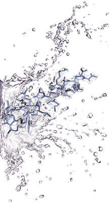
A team from the Carragher lab has imaged a protein complex at the highest resolution ever achieved with single particle cryo-electron microscopy. The image reveals individual molecules at 2.8 Å and is, to the researchers’ knowledge, the first published research using this technique that shows individual water molecules.