


- VOL 14. ISSUE 22
August 11, 2014
SEARCH NEWS & VIEWS

Team Finds New Calorie-Burning Switch in Brown Fat
Scientists Create Most Detailed Map Yet of Virus that Causes Colds, Pink Eye
Researchers Find Genetic Mutations Linked to Salivary Gland Tumors
TSRI Scientists Awarded $3.4 Million to Investigate the Secret Lives of Brain Cells
NEWS & VIEWS HOME
PAST ISSUES
KUDOS
SCIENTIFIC CALENDAR
CA AUDITORIUM EVENTS
CONTACT

- Laboratory Equipment
Scientists Discover Clues to Altered Brain Wiring in Autism - The San Diego Union-Tribune
Our perpetually vigilant internal guardian


FOLLOW US


Scientists Create Most Detailed Map Yet of Virus that Causes Colds, Pink Eye
By Madeline McCurry-Schmidt
Meet the adenoviruses, some of the most common viruses around. Is your coworker coughing? Could be an adenovirus. Your toddler have pink eye? Hello, adenovirus.
Though they usually just cause minor infections, adenoviruses can turn deadly in people with compromised immune systems, such as those receiving chemotherapy or organ transplants. The viruses are also of interest for their vaccine and gene-delivering potential in new therapies to treat diseases such as cystic fibrosis.
At The Scripps Research Institute (TSRI), researchers Vijay Reddy and Glen Nemerow have now created the most detailed map yet of the adenovirus—the largest virus structure ever solved to this degree of detail. The work, published recently in the Early Edition of the journal Proceedings of the National Academy of Sciences, advances the understanding of the basic features of the virus and possible ways to stop it.
New insights from the work include the discovery of a unique protein that busts through host cell membranes “like a sledgehammer.” The research also shows for the first time the positions of “cement” proteins that bind the parts of the virus’s outer shell and improves previous models.
A Decades-Long Mystery
For years, Reddy and Nemerow had been curious about the structure of adenoviruses. In a 2010 study, the researchers mapped out major parts of the structure, but they knew they weren’t seeing everything.
“We had pretty interesting information, but the story was not complete,” said Reddy.
Also in 2010, researchers from a laboratory at the University of California, Los Angeles, published what they believed to be a more complete structure of an adenovirus, but this structure did not explain mysterious data that scientists had been collecting since the 1970s. “That’s one thing we worried about—the previous model was not fully consistent with the experimental data,” Reddy said.
Reddy and Nemerow aimed to get a more detailed look at this structure, but first they had to jump some hurdles. To map the structure in three dimensions, the team used X-ray crystallography, a technique to determine the structure of proteins in crystals. It was tricky, however, to get the proteins in the virus to align into useful crystals—a challenge that had held back their 2010 study. Then they had to collect and process the data and build maps to show the positions of the atoms in the proteins.
Reddy said the project, which they began in 1998, demanded persistence. The researchers had to try new procedures and scour scientific journals to explain how the virus’s structure functioned.
Their persistence paid off. In a paradigm shift, the new study shows that some proteins thought to be inside the virus’s shell are actually on the outside. In addition, the researchers spotted two new proteins that had never been seen in adenoviruses before. They compared the positions of these proteins to what they knew about the function of the virus, and they realized that their revised structure explained the data from the 1970s.
Nemerow said these kinds of corrections are common in the field. As technology improves and scientists try new methods, they can see more details in a structure.
“This is not unheard of in structural biology,” said Nemerow. “We had a deeper knowledge of the biochemistry and the function of the virus, and that helped in our model building.”
A Powerful Protein
The new detailed picture of the adenovirus structure shows a spherical shell made up of capsid proteins and held together by cement proteins. A newly discovered protein, called protein VI, attaches to several spots on the inside of this shell.
Nemerow compares protein VI to a sledgehammer, using brute force to break through a host cell’s membrane so viral DNA can be inserted into the host cell, which then becomes a virus-producing factory.
If scientists can disrupt the action of protein VI, they might be able to stop adenovirus infections, said Reddy. There is also the possibility of disrupting the cement proteins that hold the virus together. “If you eliminate the cement proteins, then the virus does not form,” said Reddy.
With the refined structure in hand, scientists may also be able to harness the activity of protein VI for gene therapy. Instead of delivering harmful genes, the virus could deliver genes to treat genetic diseases. “It’s possible that gene transfer and antivirals could evolve from better knowledge of the virus structure—and protein VI in particular,” said Nemerow.
The Next Steps
By bringing structural biology and immunology together, Reddy and Nemerow were able to advance biomedical knowledge. “This wouldn’t have happened outside the Scripps environment,” said Reddy.
“It’s one of the most exciting collaborations I’ve had since I’ve been at Scripps, and there’s still more work to be done,” said Nemerow.
The next steps are to study how the immune system interacts with adenoviruses and to map out the locations and structures of minor components within the viruses. Further research could explain why some adenoviruses attack the eyes, for example, while others attack the respiratory system.
Funding for the research was provided by the National Institutes of Health National Institutes of Health (AI070771, AI103692, HL054352, Y1-CO-1020, Y1-GM-11040) and the US Department of Energy (DE-AC02-06CH11357).
Send comments to: press[at]scripps.edu
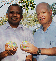
“[This is] one of the most exciting collaborations I’ve had since I’ve been at Scripps, and there’s still more work to be done,” says Professor Glenn Nemerow, shown here with long-time collaborator Associate Professor Vijay Reddy. (Photo by Cindy Brauer.)
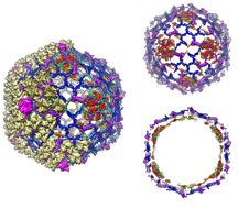
This image shows the organization of adenovirus cement proteins with respect to major capsid proteins. Click for details. (Image by Vijay Reddy and Glen Nemerow.)