


- VOL 14. ISSUE 20
July 14, 2014
SEARCH NEWS & VIEWS

State-of-the-Art Electron Microscope Opens New Vistas for TSRI Researchers
Team Sheds New Light on Nerve Cell Growth
Scientists Uncover New Compounds that Could Affect Circadian Rhythm
In Memoriam: Janet R. “Jean” Kellogg (1917–2014)
Researchers Awarded $2.6 Million to Develop Stem Cell Lines for Global Research
NEWS & VIEWS HOME
PAST ISSUES
KUDOS
SCIENTIFIC CALENDAR
CA AUDITORIUM EVENTS
CONTACT

- Laboratory Equipment
Scientists Discover Clues to Altered Brain Wiring in Autism - The San Diego Union-Tribune
Our perpetually vigilant internal guardian


FOLLOW US


State-of-the-Art Electron Microscopes Open New Vistas for TSRI Researchers
By Madeline McCurry-Schmidt
Twice this year, a truck pulled up at the La Jolla campus of The Scripps Research Institute (TSRI) and a small crowd of faculty and staff watched while a series of wooden crates were wheeled gingerly off the back.
These special deliveries were the Titan Krios and the Talos, two massive transmission electron microscopes (TEMs) built by the FEI Company. Already world leaders in electron microscopy, researchers at TSRI are using these new microscopes as part of an advanced microscopy suite to study molecules involved in diseases from cancer to AIDS.
The Titan Krios and the Talos TEMs, which were installed in a custom-designed suite, allow researchers to capture three-dimensional microscopy images that are higher-resolution than ever before.
“We’re not limited by technology anymore,” said Bridget Carragher, professor at TSRI and co-director of the National Resource for Automated Molecular Microscopy. “It’s a big step. We’re very, very happy.”
A Closer Look at Disease
As cryo-electron microscopes, the Titan Krios and Talos examine samples cooled to liquid nitrogen temperatures and illuminate them using high-energy electrons. The electrons beam down through a vertical, tube-like microscope column, and electro-magnetic lenses focus the beam of electrons to capture a high-resolution image of the sample.
Carragher compared the Titan Krios to a new pair of eyeglasses—in this case the most powerful eyeglasses currently available.
The Talos is also unique—the Talos at TSRI is the first Talos instrument to be installed anywhere in the world at a customer site. “We are impressed by the stability and reliability of this brand new instrument and optimistic that it will provide us with an outstanding solution intermediate between our high-end Krios and our more standard workhorse instruments,” said Carragher.
Andrew Ward, assistant professor at TRSI, and his lab have begun to use the Titan Krios and the Talos to better understand a major health problem: HIV. Dmitry Lyumkis, a research associate who recently earned his PhD in Carragher’s lab at TSRI, worked with Ward on the HIV project using TSRI’s other electron microscopes to determine the structure of a three-part protein, called the Envelope trimer, which sticks out of the surface of the HIV virus.
Understanding the trimer’s structure is key to developing an HIV vaccine, and images from the Titan Krios will help researchers see new parts of the virus that interact with the human immune system. “It will get us better resolution,” said Lyumkis.
Anchi Cheng, a senior staff scientist in Carragher’s lab, worked long hours to set up the Titan Krios and Talos as soon as they arrived. She looks forward to using the microscopes to visualize the finest details of protein macromolecules, including the amino acid peptide side-chains. So far, she has only studied side-chains at a resolution of 4.5 Angstroms (one ten-billionth of a meter).
“Because of its stability and better optics, we expect to be more routinely achieve resolution in the 3 to 4 Angstrom range, making viewing of all peptide side-chains much easier,” said Cheng.
Better Tools for Research
The Titan Krios provides better images because it solves several problems that limits other microscopes.
First, the Titan Krios uses a higher energy electron source—300 Kiloelectron-volts (KeV), compared to 200KeV on all the other TEMs available. This allows researchers at TSRI to examine much thicker specimens and understand their three-dimensional structure. The Titan Krios also has an additional electro-magnetic lens that provides a highly parallel electron beam and results in higher-resolution images.
The technology behind the Titan Krios also solves a problem with stability. Typical electron microscopes are vulnerable to the smallest vibrations—from the slightest breeze to the sound of a person speaking. The Titan Krios solves this problem by housing the entire system in a 12-foot-tall protective case.
Many electron microscopes also require researchers to cool samples by manually adding liquid nitrogen to the system every few hours. But the Titan Krios has automated cooling and specimen-changing systems, which speed up studies and give more researchers time to use the equipment.
The Titan Krios is coupled to a new generation of digital camera, the Gatan K2 Summit, which detects electrons directly instead of having to convert electrons to light. This provides for much higher resolution images. The new detectors also acquire images at a very high speed, essentially capturing a movie of the specimen being imaged, rather than a single frame. This allows any movement the specimen makes during imaging to be corrected with even further and dramatic improvements to the resolution.
Though the Talos does not have all of these advanced features, it does have improved stability and a Piezo stage, which allows for very precise sample positioning.
Teamwork at TSRI
Like many projects at TSRI, capturing and analyzing data from these microscopes is a team effort.
Installing the Titan Krios started with the TSRI facilities staff, who designed and built a special suite to control vibrations, temperature, air flow and humidity. This unique room is just steps away from the room that houses the Talos.
“This is probably one of the best microscope suites in the world,” said Clint Potter, professor at TSRI and co-director of the National Resource for Automated Molecular Microscopy.
The TEM and the Titan Krios also take advantage of software created by Potter and Carragher to automatically control the microscope and acquire images of the specimen of interest. This software helps scientists save time and greatly increases the number of samples and number of images of each sample that can be routinely acquired.
Once scientists have images from these microscopes, they make extensive use of TSRI computational services for analysis. “We have great computational facilities and really outstanding computational staff that make this possible,” said Carragher.
In addition to breaking new scientific ground, Potter said working with the Titan Krios gives TSRI graduate students and postdoctoral fellows a chance to learn how to use automated electron microscopy equipment, work with imaging software and try new data collection methods.
“They’re collaborating with the top biological people at Scripps,” said Potter. “That makes you a hot commodity.”
Time on the Titan Krios and Talos TEM is shared among the electron microscopy laboratories at TSRI, led by Potter, Carragher, Andrew Ward, Ian Wilson, Jack Johnson, Francisco Asturias, Ronald Milligan, Gabriel Lander and Nigel Unwin, who are using the microscope for a range of projects, including studies of molecules involved in disease and those critical to human health. Funding for the new microscopes largely came from National Institute of Health’s National Institute of Allergy and Infectious Diseases, through a supplement to the Scripps Center for HIV/AIDS Vaccine Immunology and Immunogen Discovery (UM1 AI100663); the Bill & Melinda Gates Foundation; and TSRI.
Send comments to: press[at]scripps.edu
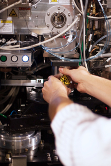
“This is probably one of the best microscope suites in the world,” says Clint Potter, professor at TSRI and co-director of the National Resource for Automated Molecular Microscopy. Shown above is part of the new Titan Krios.(Photo by John Dole.)