


- VOL 13. ISSUE 33
November 04, 2013
SEARCH NEWS & VIEWS

Scientists Capture Most Detailed Picture Yet of Key AIDS Protein
Preclinical Study Offers Hope for New Approach to Controlling HIV
NIH Names George Koob Director of the National Institute on Alcohol Abuse and Alcoholism
NEWS & VIEWS HOME
PAST ISSUES
KUDOS
SCIENTIFIC CALENDAR
CA AUDITORIUM EVENTS
CONTACT

- Laboratory Equipment
Scientists Discover Clues to Altered Brain Wiring in Autism - The San Diego Union-Tribune
Our perpetually vigilant internal guardian


FOLLOW US


Scientists Capture Most Detailed Picture Yet of Key AIDS Protein
Collaborating scientists at The Scripps Research Institute (TSRI) and Weill Cornell Medical College have determined the first atomic-level structure of the tripartite HIV envelope protein—long considered one of the most difficult targets in structural biology and of great value for medical science.
The new data provide the most detailed picture yet of the AIDS-causing virus’s complex envelope, including sites that future vaccines will try to mimic to elicit a protective immune response.
“Most of the prior structural studies of this envelope complex focused on individual subunits, but we’ve needed the structure of the full complex to properly define the sites of vulnerability that could be targeted, for example with a vaccine,” said Ian A. Wilson, the Hansen Professor of Structural Biology at TSRI, and a senior author of the new research with biologists Andrew Ward and Bridget Carragher of TSRI and John Moore of Weill Cornell.
The findings are published in two papers in Science Express, the early online edition of the journal Science, on October 31, 2013.
A Difficult Target
HIV, the human immunodeficiency virus, currently infects about 34 million people globally, 10 percent of whom are children, according to World Health Organization estimates. Although antiviral drugs are now used to manage many HIV infections, especially in developed countries, scientists have long sought a vaccine that can prevent new infections and would help perhaps to ultimately eradicate the virus from the human population.
However, none of the HIV vaccines tested so far has come close to providing adequate protection. This failure is due largely to the challenges posed by HIV’s envelope protein, known to virologists as Env.
HIV’s Env is not a single, simple protein but rather a “trimer” made of three identical, loosely connected structures with a stalk-like subunit, gp41, and a cap-like region, gp120. Each trimer resembles a mushroom and up to about 15 of these Env trimers sprout from the membrane of a typical virus particle, ready to latch onto susceptible human cells and facilitate viral entry.
Although Env in principle is exposed to the immune system, in practice it has evolved highly effective strategies for evading immune attack. It frequently mutates its outermost “variable loop” regions, for example, and also coats its surfaces with hard-to-grip sugar molecules (also called glycans).
Even so, HIV vaccine designers might have succeeded by now, had they been able to study the structure of the entire Env protein at atomic-scale—in particular, to fully characterize the sites where the most effective virus-neutralizing antibodies bind. But Env’s structure is so complex and delicate that scientists have had great difficulty obtaining the protein in a form that is suitable for atomic-resolution imaging.
“It tends to fall apart, for example, even when it’s on the surface of the virus, so to study it we have to engineer it to be more stable,” said Ward, who is an assistant professor in TSRI’s Department of Integrative Structural and Computational Biology.
The key goal in this area has been to engineer a version of the Env trimer that has the stability and other properties needed for atomic-resolution imaging, yet retains virtually all the quaternary structure found on native Env.
Illuminating Infection
After many years in pursuit of this goal, Moore, Rogier W. Sanders and their colleagues at Weill Cornell Medical College, working with Wilson, Ward and others at TSRI, recently managed to produce a version of the Env trimer called BG505 SOSIP.664 gp140 that is suitable for atomic-level imaging work—and includes all of the trimer structure that normally sits outside the viral membrane. The TSRI researchers then evaluated the new Env trimer using advanced versions of two imaging methods, X-ray crystallography and electron microscopy. The X-ray crystallography study was the first ever of an Env trimer, and both methods resolved the trimer structure to a finer level of detail than has been reported before.
“The new data are consistent with the findings on Env subunits over the last 15 years, but also have enabled us to explain many prior observations about HIV in structural terms for the first time,” said Jean-Philippe Julien, a senior research associate in the Wilson laboratory at TSRI, who was first author of the X-ray crystallography study.
The data illuminated the complex process by which the Env trimer assembles and later undergoes radical shape changes during infection and clarified how it compares to envelope proteins on other dangerous viruses, such as flu and Ebola.
Arguably the most important implications of the new findings are for HIV vaccine design. In both of the new studies, Env trimers were imaged while bound to broadly neutralizing antibodies against HIV. Such antibodies, isolated from naturally infected patients, are the very rare ones that somehow bind to Env in a way that blocks the infectivity of a high proportion of HIV strains.
Ideally an HIV vaccine would elicit large numbers of such antibodies from patients, and to achieve that, vaccine designers would like to know the precise structural details of the sites where these antibodies bind to the virus—so that they can mimic those viral “epitopes” with the vaccine.
“It has been a privilege for us to work with the Scripps team on this project,” said Moore on behalf of the Weill Cornell group. “Now we all need to harness this new knowledge to design and test next-generation trimers and see if we can induce the broadly active neutralizing antibodies an effective vaccine is going to need.”
“One surprise from this study was the revelation of the complexity and the relative inaccessibility of these neutralizing epitopes,” said Julien. “It helps to know this for future vaccine design, but it also makes it clear why previous structure-based HIV vaccines have had so little success.”
“We found that these neutralizing epitopes encompass features such as the variable loop regions and glycans that were excluded from previous studies of individual Env subunits,” said Dmitry Lyumkis, first author of the electron microscopy study, who is a graduate student at TSRI participating in the NIH-funded National Resource for Automated Molecular Microscopy. “We observed, too, that neutralizing antibody binding to gp120 can be influenced by the neighboring gp120 structure within the trimer—another complication that was not apparent when we were not studying the whole trimer.”
Having provided these valuable structural insights, the new Env trimer is now being put to work in vaccine development. “We aim to refine and increase the antibody response, then move forward to tests in humans,” said Ward.
Other contributors to the studies, “Cryo-EM structure of a fully glycosylated soluble cleaved HIV-1 envelope trimer,” and “Crystal structure of a soluble cleaved HIV-1 envelope trimer,” included TSRI’s Natalia de Val, Devin Sok, Robyn L. Stanfield and Marc C. Deller; and Weill Cornell Medical College’s Rogier W. Sanders (also at Academic Medical Center, Amsterdam), Albert Cupo and Per-Johan Klasse. In addition to Wilson, Ward and Carragher, senior participants at TSRI included Clinton S. Potter and Dennis Burton.
The research was supported in part by the National Institutes of Health (HIVRAD P01 AI82362, CHAVI-ID UM1 AI100663, R01 AI84817, R37 AI36082, R01 AI33292), the US NIH NIGMS Biomedical Research Technology Program (GM103310), and the International AIDS Vaccine Initiative Neutralizing Antibody Consortium and Center.
For more information on the papers, see http://www.sciencemag.org/content/early/2013/10/30/science.1245627.abstract and http://www.sciencemag.org/content/early/2013/10/30/science.1245625.abstract
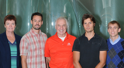
The TSRI researchers behind the new HIV findings include (left to right) Bridget Carragher, Jean-Philippe Julien, Ian Wilson, Dmitry Lyumkis and Andrew Ward. (Photo by Dave Freeman.)
Send comments to: press[at]scripps.edu

Using advanced techniques in both cryo-EM and x-ray crystallography, a team has now determined the structure of the HIV envelope protein—long considered one of the most difficult targets in structural biology and of great value for medical science. Here, the protein is shown bound by broadly neutralizing antibodies against two distinct sites of vulnerability.

A video close up of the HIV envelope protein.