|
(Page 2 of 2)
"What we did was to take this peptide that we knew could
inhibit the binding and infection of measles and display it
on the surface of the plant virus," says Manchester.
What Manchester and Johnson found was that not only were
they able to protect cells from measles infection, but they
were able to do so with at least 100-fold greater efficacy
than with the peptide alone, and Manchester was able to prevent
infection in vivo. The secret of the CPMV's success,
they believe, is its polyvalency—the fact that it displays
multiple copies of the anti-measles peptide.
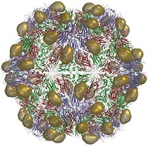
"When the [cowpea mosaic] virus comes in contact with the
measles virus, it's not just bringing one copy, but 60 copies,"
says Johnson. Moreover, the hemagglutinin molecule to which
the peptides bind is a trimer and so binding is favorable.
And they could potentially attach more than one peptide to
the cow pea mosaic virus and target measles with even higher
efficacy.
The same approach might be used to create a vaccine in the
traditional sense by putting antigen molecules from measles
or some other virus that would stimulate an immune response
to block an infection from a later exposure.
One of the great advantages of using such an approach is
its frugality—the virus does the work of making the peptide.
This is an advantage for achieving Manchester's ultimate goal
of making new vaccines, since one of the main criteria for
any globally effective vaccine is its price.
Another preferential feature for a vaccine's success is
its bioavailability, since delivering a vaccine to remote
regions is made much more difficult if specialized equipment
or training is required. And Manchester says that the modified
CPMV looks reasonably orally bioavailable, based on some preliminary
studies she completed with Finn.
Finn attached fluorescent dyes to the viral particles to
see where they go in vivo and how long they last in
tissues, and Manchester found that, indeed, the CPMV molecules
are distributed throughout the organism. Now she and Finn
are trying to make further improvements to this distribution
by attaching polyethylene glycol molecules to the virions
to dampen the immune response to them.
"If you coat a protein with polyethylene glycol, it tends
to dramatically reduce its visibility to the immune system,"
says Johnson.
Bringing their Collaboration to the New Center
Finn, Johnson, Lin and Manchester are all part of the new
Center for Integrative Molecular Biosciences (CIMBio) faculty
and have laboratory space in the newly constructed CarrAmerica
B building. CIMBio was designed to be the most advanced biological
microscopy center in the world and to provide an environment
in which the expertise and resources of many research groups
could be combined.
Finn and Johnson provide a service to the center by providing
tailored CPMV for the molecular microscopes and for the new
automated processes. Since the structure is so well determined,
it makes a good test bed to determine how well the electron
microscopes are doing.
Finn will also conduct an independent program on basic research
into new labels for electron microscopy. In EM, heavy atom
labels are routinely attached to the particular molecules
of interest in order to image these molecules. Currently,
there are only a few commercially available labels.
"That's just not versatile enough for the kinds of applications
this center is going to deal with," says Finn, adding that
the result of his research into new labels will be a specialized
chemistry service for researchers using the CIMBio facilities.
"We want biologists and biochemists to come to us with a
problem, 'Here's a protein I need to label with a heavy atom
residue' or 'I tried what's available and it doesn't work
so well.' That's partly what we're here to do."
For the most part, though, Johnson, Lin, Finn, and Manchester
are collaborating with each other to find and test uses for
the modified CPMV particles. And they have many possibilities.
They have already successfully attached biotin (Vitamin
B), sugars, and organic chemicals to the viral surface, and
they can immobilize large molecules on the surface—whole
proteins even.
"We can attach anything we want to the surface of the virus,"
says Johnson.
One possible attachment are molecules that can be used to
image cancers or other biological states in living cells by
labeling CPMV with an anti-tumor agent or some molecule that
targets a particular biology of interest along with radiolabels
or some other sensing agent that could be visible under magnetic
resonance or microscope imaging.
Finn, Johnson, and Lin found that cysteines could also be
double-labeled by placing cysteine on both the inside and
outside of the virus shell and that a pattern of attachment
sites could then be created that would allow for novel chemistry.
Catalysis could potentially be carried out with the virus
particles. The temperature and pH stability, solubility, and
the chromatographic properties of the virus can be altered
at will, by adding the right molecules. And the virus particles
can self-organize into network arrays in a crystal, which
may make it a useful building block for various applications
in nanotechnology, the field that seeks to build functional
material on the nanometer scale (roughly one to one hundred
billionths of a meter).
"You can, in principle, determine the type of assembly you
get by programming the building blocks," says Finn.
And in collaboration with Manchester, Finn and Johnson are
excited about the possibility of testing CPMV as a polyvalent
delivery vehicle. Since there are 60 attachment sites, the
virus will present multiple copies of the attached molecules
wherever it goes.
"This is something uniquely Scripps," says Johnson. "We
have three different departments, three different backgrounds,
and yet here we are."
1 | 2 |

|