|
(page 2 of 2)
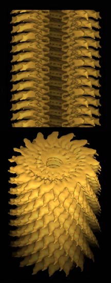
A single high-resolution image of a sample under an electron
microscope has too much noise to yield accurate molecular
representation. Images must be averaged together with their
counterparts to reduce noise. Plus, any single molecular assembly
imaged will be but one 2-D projection of what is a 3-D object,
so the averaging must be done over many possible angles. To
build a 3-D model, one must take many images and build a structure
by looking at all the different angles of all the different
molecular assemblies imaged.
Building a 3-D model is like looking at a piece of sculpture
in a gallery. Only by walking around the piece and viewing
its various sides and angles can the brain build a mental
image of the art and fully comprehend its dimension, perspective,
and scale. The same is true using a computer. Only by piecing
together many different views of a molecule from a microscope
can a computer build a model of the molecular assembly.
And the molecule that is being imaged gets destroyed in
the process, so the next image must be captured from some
other part of the sample holder grid. This has always required
a person to choose different spots on the grid manually. As
the number of grid spots goes up, so goes the level of tedium.
"What we really want is 100,000 to 1,000,000 molecule images
and that just takes too long to do manually," says Carragher.
"Then you want to do 10 different conformational states, 20
different labeling studies, and each time it's going to take
three to six months. That's more than the lifetime of a graduate
student."
"There are projects," Carragher adds, "projects people just
don't do because the manual labor required is just too daunting."
Carragher and Potter, who lead the Automated Molecular Imaging
group, are creating algorithms for automated data collection
and analysis, which should simplify the technique of electron
microscopy and enable throughput to be increased dramatically.
So Long, John Henry
Several years ago, Carragher and Potter suggested that automated
data collection and analysis could be developed for EM. A
similar goal had been accomplished in x-ray crystallography,
and given the need for structural information in our post-genomics
proteomics world, automation would represent significant progress.
So Carragher and Potter started developing automated EM
algorithms and began writing grants with Milligan to develop
these into programs. "It took off from there," says Carragher.
They succeeded in developing software for both the collection
and the analysis, which they brought to TSRI when they came
last year to form the Automated Molecular Imaging group at
TSRI. Milligan helped recruit his long-time collaborators
from the University of Illinois at Urbana–Champaign,
where they were co-directors of the Imaging Technology Group
of the Beckman Institute for Advanced Science and Technology.
Creating the algorithms was not easy. Using the manual technique,
a person has to make decisions about where to focus the EM
beam and take a picture, looking first at low resolution and
then deciding in which areas to collect data at high resolution.
For automation to succeed, the computer must do the same thing
and use intelligent criteria to search the low resolution
image for appropriate targets.
"Even a two-year-old can tell a cat from a dog, but that's
a very hard problem for a machine," says Carragher. "But what
humans are not good at is doing the same boring thing a thousand
times in the dark for weeks."
Carragher and Potter had to write their software to take
a low-resolution image, select areas to image in medium resolution,
and then analyze that image and strip out targets for high-resolution
maps. Then, they had the computers put the data into processing
programs and calculate 3-D maps. Recently, they have been
testing and refining the programs.
"What we have done over the past year is to show that you
can insert a [sample] in the microscope and [calculate] a
3-D map fully automatically," says Potter.
In fact, Carragher and Potter constructed one of the best
3-D maps of the tobacco mosaic virus in under two days. By
comparison, the work would have taken several months of work
just a few years ago and perhaps several weeks using conventional
methods today.
"We can now go from inserting a specimen in the microscope
to having a 3-D map in 24 hours," says Milligan, adding that
the fear of failure should no longer be a limiting factor
for experiments.
Still, the automation is not fully implemented, so one of
the immediate goals of the Automated Molecular Imaging Group
is to see their software used for practical applications,
something that their coming to TSRI will facilitate.
"There are so many people who want to collaborate with us
here—it's great," says Potter, adding that within a few
months of their arrival they had already found an almost overwhelming
number of projects.
"At the moment we need to make the technique very efficient
and very general, [and] get it out to the community" says
Carragher. "We can do it, and now we want to be able to do
it routinely for anybody."
Additional plans include the design of technology that would
make EM high-throughput. This includes a robotic specimen
handler that Carragher and Potter have been experimenting
with that would allow the instruments to be left alone to
collect and analyze even larger sets of data.
"You could look at maybe 10 grids overnight," says Potter.
Ready for Tours
Though the shared space of the CarrAmerica B building and
the collaborations it fosters within CIMBio and throughout
the TSRI campus will be reward enough, there is one more thing
that the building provides: ready-made tours.
In the plans that Milligan and the others drew up, they
envisioned several people controlling the microscopes and
discussing the images as they are collecting data. They also
anticipated people peering through the glass wall of the control
room, and this has its advantages.
"It's a very easy way to communicate what we are doing,"
Carragher says to me as we walk past the control room on a
recent tour of the facilities. A technician had one of the
new microscope's back panels open and was busy fiddling with
some wires.
"Is it done yet?" Carragher asks.
1 | 2 |

|