Almost There:
Cutting-Edge Molecular Microscopy Center Prepares to Open
By Jason Socrates
Bardi
"It
is very easy to answer many of these fundamental biological
questions; you just look at the thing!... Make the microscope
one hundred times more powerful, and many problems of biology
would be made very much easier. I exaggerate, of course,
but the biologists would surely be very thankful to you—and
they would prefer that to the criticism that they should
use more mathematics."
——Richard
P. Feynman. From There's Plenty of Room at the Bottom,
a lecture given to the American Physical Society in 1959.
When Associate Professors Bridget Carragher and Clint Potter
arrived at The Scripps Research Institute (TSRI) last year,
they knew where their laboratory space would eventually be,
but they had no idea what the space would be like. That is,
until after they sat down with Professor Ron Milligan over
drinks one night and drew up plans on a blank blueprint of
the interior of the CarrAmerica B building.
In the following weeks, this became the blueprint for Milligan's
dream of the most advanced biological microscopy center in
the world—the Center for Integrative Molecular Biosciences
(CIMBio)—which officially opens next month. CIMBio is
built around its advanced microscopes and open laboratories
and houses several TSRI faculty under one roof.
"We had an almost unique opportunity to design the best
electron microscopy suite, and we put a lot of effort into
doing this," says Milligan.
The design is predicated on six rooms for microscopes, which
are at the center of the building. The microscopes are mounted
on three-foot-thick concrete slabs isolated from the building's
foundation, which protect the instrumentation from vibrations.
The rooms are climate-controlled with low humidity to prevent
contamination of samples by water vapor, and they are sound-proofed
so that noise from the corridors does not cause vibrations.
The air supply coming into the rooms passes through a nylon
sleeve that breaks up any air currents, and the microscopes
can be controlled entirely from a separate room so that the
samples can be left alone in the dark under the microscopes.
"It's quiet, there are no air currents, and the microscopes
are sitting on a very stable platform," says Milligan.
Ground broke on the interior design of CarrAmerica B in
March, and the construction lasted throughout the fall. The
first groups moved in at the end of December. A year ago,
CarrAmerica B was a shell. Now it is an oyster with more than
one pearl.
Molecular Machine Mania
Milligan, Carragher, and Potter are all founding members
of the CIMBio, which was organized to combine the talents
of several groups across campus.
The center seeks to speedily obtain and analyze high-resolution
structural images of large molecular complexes of the cell
by combining the use of x-ray crystallography and electron
microscopy (EM). CIMBio members include investigators Francisco
Asturias, M.G. Finn, Jack Johnson, Elizabeth Wilson-Kubalek,
Mari Manchester, Nigel Unwin, and Mark Yeager.
What unites the members of CIMBio is their interest in the
combined use of the x-ray crystallography and EM techniques
as a means to unravel the structure and mechanism of action
of the large molecular assemblies of the cell—such as
the transcription complexes that make messages from the genes,
membrane channels and pumps that import and export materials,
and the tiny molecular tracks and motors that move cells and
form important structures like the mitotic spindle.
Phase I of CIMBio will be devoted to working out the structure
of the proteins and nucleic acids in complexes that carry
out the work of the cell.
While the individual protein components of these machines
may be studied by x-ray crystallography, the machines themselves
are compositionally and conformationally dynamic, making them
unsuitable for x-ray methods. They are, however, ideal specimens
for electron microscopy. Polymerases, membrane complexes,
viruses, and motor proteins can all be visualized in their
native environment using EM.
Phase II will concentrate on the dynamics of those cellular
machines—their assembly, disassembly, and control over
time.
Laboratory space for that effort is already under construction
in CarrAmerica B, and at the end of the year, investigators
Velia Fowler, Klaus Hahn, Clare Waterman-Storer and Kevin
Sullivan will relocate there to lead the Phase II efforts.
The building combines several of these laboratories into
one contiguous shared space built above and around the microscopes.
The laboratories have an open design and some of the facilities—like
the microscopes and an imaging area—are shared, something
that the CIMBio researchers appreciate.
"This is a collection of widely diverse scientists, and
we want to maintain and enrich our collaborations" says investigator
M.G. Finn, whose group was the first to move into the new
space. "Here we can't help running into each other."
EM Imaging of Biological Structures
Electron microscopy, which has been around since the 1930s,
uses a beam of electrons to image tiny objects onto a digital
camera or a photographic plate. EM looks at a range of magnifications,
from no more than an ordinary microscope that magnifies up
to 60 times to those that magnify up to 1,000,000 times. CryoEM,
which is the technique used for viewing biological materials,
requires the samples to be spread to a thin film and frozen
on a copper meshwork grid.
The final products of these electron images are 3-D maps,
which are representations of the cellular structures on the
slide at near-atomic resolutions—up to about 3-4 angstroms
under the best of circumstances. When combined with the x-ray
structures of the component parts of the structures, EM maps
can yield a detailed description of the structure and action
of the entire machine.
Further application of this technique will be an invaluable
tool for studying membrane-bound proteins, which are notoriously
hard to crystallize. Less than one half of one percent of
the structures contained in the Brookhaven National Laboratory
Protein Data Bank are of integral membrane proteins, despite
the fact that over a third of all proteins in the body are
in the membrane.
But EM is not a routine technique. Calculating an EM structure
manually takes weeks or even months. It can be tedious.
1 | 2 |

|
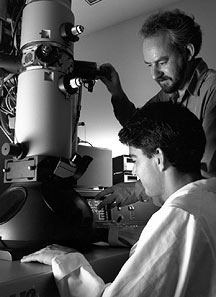
Professor Ron Milligan had a dream of
building the most advanced biological microscopy center in
the world. Milligan, standing, is seen here working with MCSC
graduate James D. Jontes. Photo by Michael
Balderas.
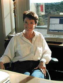
Associate Professor Bridget Carragher
(above) is working with Associate Professor Clint Potter to
create algorithms for automated data collection and analysis,
which should simplify the technique of electron microscopy
and enable throughput to be increased dramatically. Photo
by Kevin Fung.
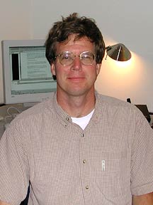
"There are so many people who want to
collaborate with us here—it's great," says Potter. Photo
by Quan Dong.
|

