Inflammation and the Brain
By Jason Socrates Bardi
"The First approached the Elephant,
And happening to fall
Against his broad and sturdy side,
At once began to bawl:
"God bless me! but the Elephant
Is very like a wall!"
The Second, feeling of the tusk,
Cried, "Ho! what have we here
So very round and smooth and sharp?
To me 'tis mighty clear
This wonder of an Elephant
Is very like a spear!"
The Third approached the animal,
And happening to take
The squirming trunk within his hands,
Thus boldly up and spake:
"I see," quoth he, "the Elephant
Is very like a snake!"
—John Godfrey Saxe, Blind Men & An Elephant
Most people know the allegory in which several blind men argue over the nature of the elephant. Infuriatingly, all are right in their descriptions of the creature, but all describe only a single part. None can give a truly comprehensive picture of the beast.
A similar problem confronts scientists today who attempt to understand and describe complex brain-related phenomena—such as the causes of addiction and craving, the consequences of brain infections and disease, and the effects of inflammation in the brain. The brain, like the elephant, does not easily yield to reductionism.
Understanding such complicated brain-related phenomena requires assembling and analyzing details on different levels—individual molecules, groups of cells, whole tissues and organs, and an organism’s individual and group behavior. Asking a question like, what is the effect of substance X on the brain? will likely yield a diverse set of answers, depending on which approach a scientist takes.
“The thing about the brain is you can’t do a [single] specific experiment,” says Donna Gruol, who is an associate professor in the Department of Neuropharmacology at The Scripps Research Institute. “In order to understand how things affect the brain, you have to understand it on many levels—from the behavioral down to the molecular—and be able to integrate all these levels.”
Gruol is an electrophysiologist. She studies the electrical activity of neurons—how ion channels on the neurons work to let charged particles in and out of a cell and how neurons propagate such a signal to its axon where it communicates over the synapse with another neuron. She looks at individual neurons and works out how they communicate with each other in different parts of the brain, and how those parts of the brain interact with other parts of the brain.
The Importance and Difficulty of Studying Neurons
Gruol joined what would eventually become today’s Scripps Research Department of Neuropharmacology in 1979, when she became a senior research associate in Floyd Bloom’s group at the Salk Institute for Biological Studies. When Bloom came to Scripps Research a few years later, Gruol followed and established her own laboratory.
In her 20-plus-year career at Scripps Research, Gruol has studied a wide range of topics, from how alcohol and drugs of abuse interact with cells of the central nervous system to the effect of an HIV infection in the brain. Through her association with the Alcohol Research Center in the Department of Neuropharmacology she currently oversees a project that is modeling binge drinking in teenagers, carried out by research associate Jilla Sabeti.
And for the last decade, she has been studying the effect of inflammation on the brain and central nervous system—questions that fall generally under the category of “neuroimmunity.”
“It’s a very new field, and it [has taken] a long time for it to grow—but it is growing,” says Gruol.
One of the reasons for the discipline’s expansion is that scientists like Gruol have been realizing the importance of inflammation in diseases and conditions that affect the brain, such as Alzheimer’s disease, head injuries, stroke, and AIDS.
“Any injury to the brain—any kind of damage or infection—causes an inflammatory response that can affect the function of the central nervous system,” she says. “The minute there is an infection in the brain, the brain cells begin producing all these [inflammatory factors].”
Address IP
One of the main inflammatory factors that Gruol studies in collaboration with assistant professor Tom Nelson and research associate Hilda Bajova has a handful of names, none of which are immediately recognizable to any average person. This chemical is variously called by its new name, the chemokine CXCL10, its old name, interferon-gamma protein 10, or simply IP-10.
IP-10 is actually a small protein of about 80 amino acids that is produced in elevated levels during a viral or bacterial infection and during the course of several other states, such as when a person suffers a traumatic blow to the head.
“We’re looking at how this chemokine affects neurons,” she says.
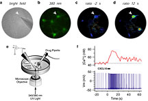
IP-10 can cause the release of intracellular calcium, which can have profound effects in the cells in which it is released. Calcium is a second messenger and regulates many different intracellular processes, including gene expression within cells.
Gruol and her colleagues have performed gene array experiments, which have revealed that some of the target genes elevated by IP-10 are other chemokines. This suggests that IP-10 can lead to the release of other inflammatory factors, which can cause even more effects in the brain.
Furthermore, Gruol recently discovered that while IP-10 can induce changes in intracellular calcium in non-neuronal cells, this effect of IP-10 occurs only in a small set of neurons. The neurons under study are in the hippocampus—a region of the brain that is critical for forming memories. Gruol treated the hippocampal neurons with IP-10 but only a few neurons responded like non-neuronal cells. Significantly, IP-10 alters another important neuronal property, neuronal excitability. This suggests that although both neurons and non-neuronal cells express receptors for IP-10, the downstream transduction pathways differ. The changes in neuronal excitability induced by IP-10 may contribute to the cognitive dysfunction associated with disease and injury in the brain.
AIDS and the Brain
Much of Gruol’s work in neuroimmunology arose initially out of her work on the effect of HIV on the brain. She was part of the Scripps AIDS Dementia Center, which directed research on the disease throughout the 1990s, and recently joined the Scripps NeuroAIDS Preclinical Studies (SNAPS) Center, a National Institute of Mental Health-supported center at Scripps Research that supports biomedical research relevant to neuroAIDS.
HIV, like other lentiviruses, has the ability to enter the central nervous system and infect cells of the brain. HIV enters the brain early in the course of infection through “Trojan horse” leukocytes—infected immune cells like macrophages, which have the ability to traffic through the brain. Once inside, the virus inside these macrophages can infect “microglia” cells, a type of non-neuronal cell in the brain.
As in other tissues in the body, HIV injures or kills these cells and spreads to infect others. And that’s only the beginning of the story.
HIV has a deleterious effect on the brain that can lead to subtle and profound neuronal dysfunction in adults. Many AIDS patients suffer from some form of central nervous system disorder in the course of their infection, ranging from minor cognitive and motor disorders to severe dementia, collectively known as neuroAIDS. In children, this is even more of an issue.
“It’s a very serious problem in pediatric AIDS,” says Gruol, adding that while some 10 to 15 percent of adult AIDS patients suffer some form of cognitive dysfunction, the prevalence is more like 50 percent among AIDS youngest victims. In these children, HIV causes cognitive malfunctions, loss of growth milestones, and behavioral problems.
The exact mechanisms through which HIV damages the brain are not known, and understanding the effect of HIV on the brain is complicated by the fact that the virus does not infect the neurons directly.
“Our hypothesis is that the virus [causes the body to produce] inflammatory factors that act on the neurons and other cells in the central nervous system,” says Gruol.
Infected macrophages and microglial cells probably overproduce chemokines and cytokines as part of the natural immune response to the HIV infection. These inflammatory factors attract immune cells, which can traffic into the brain, release more chemicals, and carry out reactions to destroy those cells that are infected with HIV. Along the way, these inflammatory factors may disrupt the function of other cells—such as neurons—that get caught in the crossfire.
The Electric Neuron
Gruol also studies issues related to the effect of one specific type of cytokine, called interleukin-6, on brain tissue and neurons. Interleukin-6 is a biological response factor produced by cells of the immune system and is linked to gene expression in immune cells.
A few years ago, Gruol discovered that interleukin-6 can change gene expression in neurons and affect neuronal function, which suggests that these immune factors can have neuroadaptive effects. Thus, while neurons themselves are not susceptible to HIV, they may be negatively affected by the chemicals produced in response to HIV infection.
Gruol and her laboratory have been able to study the actual effect of these chemicals on living neurons by using electrophysiology and calcium imaging, which makes her lab somewhat unusual in the field.
“There are very few people investigating how immune factors affect neurons using physiological approaches,” she says.
Electrophysiology measures how neurons respond to stimuli like inflammatory factors directly by using a tiny electrode to connect to and measure the conductance of a single neuron's soma (the cell body) or dendrite (the branching "process" of a neuron). Alternatively, a slightly thicker electrode can be used to measure the response of a network of neurons. Calcium imaging measures changes in intracellular calcium that occur during neuronal activity.
Regulating the excitability of neurons are different types of ion channels on neurons' surfaces. There are an array of different potassium channels, for instance, and a few sodium channels as well. These transport ions across the membrane to control the excitability of the neurons and, together with calcium channels, such important functions as the release of neurotransmitters at the synapses. To access these channels for study usually requires the use of in vitro cell culture experiments or ex vivo studies of brain tissue, both of which Gruol uses in her laboratory.
When she first started, Gruol admits, she did not know much about inflammation. “But,” she says, “I did know a lot about how to do the kind of experiments you might do to find out how inflammatory factors affect the central nervous system.”
The Whole Elephant
The lesson of the allegory of the old blind men and the elephant is clear enough: they needed only to put their piecemeal observations together to improve their understanding of the elephant as a whole.
In this sense, Gruol sees the department and the institute in general as an optimal place to study issues related to the brain because it is such a multidisciplinary place. She is able to collaborate with other scientists studying all levels of the organism—from the molecular to the behavioral. This is increasingly important as the field of neuroscience grows and our picture of the brain becomes increasingly more complicated.
Send comments to: jasonb@scripps.edu

|