$9.2 Million Grant Enables Scripps Scientists to Design
Anthrax Antitoxin Nanosponges
By Jason Socrates Bardi
A large, multi-center program project grant has been awarded
to a team of scientists at The Scripps Research Institute
(TSRI), Harvard Medical School, and The Salk Institute for
Biological Studies to discover and develop novel anthrax antitoxins
and ways of delivering them.
The overall goal of the program is to design anti-anthrax
nanosponges—antitoxin particles that could be administered
to someone who has been exposed to anthrax.
"They would basically bind up all the toxin and render it
ineffective," says TSRI Assistant Professor Marianne Manchester,
who is the principal investigator on the grant.
The "program project" grant was awarded by the National
Institute of Allergy and Infectious Diseases (NIAID) and provides
five years of funding for five projects led by investigators
at these three institutions as well as common cores that will
support the projects.
The Threat of Anthrax
Anthrax is a deadly disease that is caused by infection
with the bacterium Bacillus anthracis. It is an ancient
disease—both Homer and Virgil wrote about a disease that
was probably anthrax.
The Greeks named the disease anthrax, which means coal,
because of the characteristic black ulcers that form on the
skin of people and animals infected with the bacterium. This
cutaneous form of the disease was responsible for widespread
outbreaks among livestock through the centuries, and Louis
Pasteur famously demonstrated the first anthrax vaccine in
1881, which helped confirm the germ theory of disease.
In the 20th century, the disease and the bacterium that
causes it grew to infamy because of its potential as a biological
weapon. Over the years, several countries developed weaponized
B. anthracis spores, which cause inhalation anthrax.
B. anthracis naturally forms spores when conditions
are not right for the bacterium to replicate. When it converts
into a spore, it can lie dormant inside its protective, almost
indestructible protein coat. When spores of anthrax are breathed
in, they are taken up through the lungs by cells called macrophages.
The macrophages transport ingested spores to other parts of
the body, where they germinate into bacteria and begin reproducing
and making toxins.
Protecting against inhalation anthrax is a major public
health priority, especially after the U.S. Postal Service
attacks of late 2001. There must be an effective way to treat
individuals who have been exposed to spores as a last line
of defense.
Exposure to anthrax can be treated with antibiotics, but
the effectiveness of antibiotics diminishes over time. If
the exposure is not detected quickly enough, antibiotics alone
may not be able to save the patient. This is because B.
anthracis produces a virulent toxin that kills cells and,
in high enough doses, can kill infected people. That's the
rub—even if the infection is brought under control, the
bacteria may have produced enough toxin to be lethal.
Finding a way to neutralize the effect of the toxins would
be a great boon to public health preparedness against anthrax
exposure. That's exactly what the team on the program project
grant is trying to do.
Anthrax Toxin and Where It Binds
The anthrax "toxin" is actually a system of molecules composed
of three separate proteins released by the bacterium. Two
are virulent proteins that interact with human cells. These
are the "lethal factor," which is a metalloprotease (an enzyme
that chops up other proteins), and the "edema factor," which
is an adenylate cyclase (a protein that makes cAMP, an important
"second messenger" molecule in the body that has a variety
of systemic effects).
The third protein produced by the bacterium, called protective
antigen, is important for getting lethal factor and edema
factor into cells. Protective antigen binds to the surface
of human cells and forms a sort of cat door that allows the
lethal factor and edema factor to pass through to the interior
of the cell where they can do their damage. Once inside cells,
the lethal and edema factors lead to cell death.
The details of how the lethal and edema factors kill cells
are still somewhat murky, but what is clear is that protective
antigen, lethal factor, and edema factor work together to
make Bacillus anthracis deadly.
A few years ago, Professor John Collier of the Harvard University
Medical School and Professor John A.T. Young, who was then
at the University of Wisconsin Medical School, discovered
a human receptor of the anthrax toxin and began to elucidate
the structural details whereby anthrax toxin enters human
cells.
When protective antigen binds to these human receptors,
it inserts itself into the membrane of a cell and self-assembles
into a seven-membered heptamer, with one bound protective
antigen associating with six other identical protective antigen
proteins and forming a seven-spiked crown sticking out of
the membrane of that human cell.
This heptamer then binds to the lethal and edema factors
and acts like a pore to deliver them into the cellular membrane.
Normally, human cellular membranes—bilayers of fat, protein,
sugars, and other molecules—would normally be impenetrable
to the lethal and edema factors. But the protective antigen
heptamers enable them to pass right through the membrane and
into the cell.
Collier has had a distinguished career of studying the mechanisms
by which bacterial toxins cause disease. In the 1960s, he
showed that diphtheria toxin works by entering human cells
and inactivating an intracellular target molecule.
"As time went on, more and more bacterial toxins were found
to act inside cells," says Collier.
Anthrax toxins turn out to be one of these, and in the new
program project grant, Collier is attempting to understand
structural details of how the protective antigen binds to
human receptors on the surface of a human cell by making crystal
structures of the receptor and toxins together.
Young, who is now at the Salk Institute, has recently detected
another human receptor—called capillary morphogenesis
gene-2—to which the protective antigen also binds. In
the project he is directing, Young will be looking at the
interaction between the protective antigen and the capillary
morphogenesis gene-2 receptor, asking how these interactions
lead to the entry of the toxin.
"The discovery of a second type of anthrax toxin receptor
came as quite a surprise," says Young. "We are now attempting
to better understand how interactions between protective antigen
and both of its receptors contribute to the pathogenesis of
anthrax disease."
Collier and Young are both using this structural information
to come up with ways of inhibiting the interaction of the
protective antigen with the human receptors. In particular,
they are asking what portions of these human receptors are
necessary for the entry of the toxins and if these same parts
could be used to attach to the toxins in solution.
This is the first step in designing anti-toxins—soluble
peptides or other small molecules that could act like molecular
decoys to grab the toxins out of the bloodstream.
Nanosponges
The decoy molecules are only half the story. The other half
of is to find a good vehicle to deliver them, and that's what
Manchester and several other investigators at TSRI are working
on as their portion of the program project grant.
Peptides are more potent if they are displayed polyvalently,
which is a way of increasing the efficacy of a drug by presenting
multiple copies of it.
In fact, a few years ago, Collier identified a peptide that
was not able to inhibit the binding of protective antigen
to the human receptors on its own but could do so when it
was made in a multimeric form where one particle displayed
a score of these peptides.
"This gave us the idea that multimerization strategies could
be useful," says Manchester.
Using the targets that Collier and Young produce, the scientists
at TSRI are adapting technology that has been developed at
the institute to display these molecular decoys in multiple
numbers on the surface of nanoparticles—to make something
that they call antitoxin nanosponges.
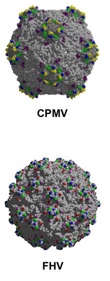
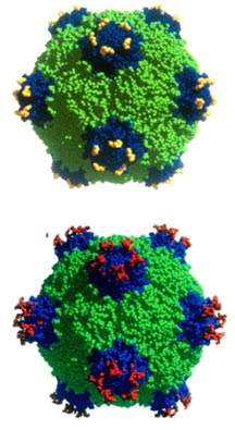
Manchester is looking at displaying the molecular decoys
on the surface of particles of Cowpea mosaic virus (CPMV).
CPMV withers and stunts the leaves and pods of the Vigna
unguiculata plant—an important crop and source of
protein in many parts of the world. Like most plant viruses,
CPMV is delivered by insects into plant cells, and like most
plant viruses, CPMV has little need for its viral envelope
to facilitate entry into cells. All these envelopes are, basically,
are rigid, stable containers—shells.
The shell of a CPMV particle is some 30 nanometers in diameter
and is formed by 60 identical copies of a pair of viral proteins
surrounding a single strand of viral RNA. These 60 pairs constitute
60 equivalent sites for displaying antitoxins.
Similarly, TSRI Associate Professor Anette Schneemann is
working with Flock house virus particles, which infect insects,
and is studying whether these will serve as an effective platform
for presenting these peptides.
The Flock house virus is about 35 nanometers in diameter
and is formed by 180 copies of a coat protein surrounding
two strands of RNA. The 180 copies each have four potential
sites for displaying an antitoxin, which means that one Flock
house virus particle could potentially display 720 copies
The structures of both of these viruses were solved a few
years ago by TSRI Professor John Johnson, who is also involved
with the grant. This means that scientists know at the atomic
level what these viruses looks like, and they can apply this
knowledge to making designer viral particles.
By changing the genetic makeup of the virus to modify the
capsid proteins, they can display peptides of interest on
the surface of the virus without altering the virus's basic
structure.
In this case, Manchester and Schneeman are taking the substances
that are produced by Collier and Young and displaying them
on the surface of their virions. Multiple copies of these
peptides can be displayed in different locations on the particle
surface, and this, they hope, will allow them to prevent the
toxin from binding to the receptor and the toxin from entering
the cells.
By Comparison
Collier and Young are also developing soluble forms of the
antitoxins that can be attached to the particle through a
chemical linker, a complementary approach to Manchester's
and Schneemann's that explores attaching the antitoxins to
the viral particles chemically .
TSRI Associate Professor M.G. Finn and Assistant Professor
Vijay Reddy, who lead this project, will compare the viral
particles to other types of scaffolds—like those made
from organic polymer or tiny flecks of gold.
"We want to be able to compare a virus to a different type
of nanoparticle to see which is more effective and why," says
Finn.
One of the advantages of the particles is that they have
the ability to traffic throughout the body and to stay in
the bloodstream for a long time. These particles are also
more stable in the gut than peptides would be, which makes
them potentially bioavailable—an important advantage
to any potential therapeutic.
The chemical or biological approaches might also be used
to create a vaccine in the traditional sense by using antigen
molecules from B. anthracis to stimulate an immune
response. This response would have the added benefit of blocking
an infection from a later exposure.
NIAID is a component of
the National Institutes of Health, an agency of the Department
of Health and Human Services. NIAID supports basic and applied
research to prevent, diagnose, and treat infectious and immune-mediated
illnesses, including HIV/AIDS and other sexually transmitted
diseases, illness from potential agents of bioterrorism, tuberculosis,
malaria, autoimmune disorders, asthma, and allergies. For
more information, see: http://www.niaid.nih.gov.
Harvard Medical School
is one of the world's preeminent institutions in medical education
and research. The breadth and depth of its scientific and
clinical disciplines are unsurpassed. The School has nearly
8,000 faculty and 17 affiliated facilities.
The Salk Institute for
Biological Studies, located in La Jolla, Calif., is an independent
nonprofit organization dedicated to fundamental discoveries
in the life sciences, the improvement of human health and
conditions, and the training of future generations of researchers.
Jonas Salk, M.D., founded the institute in 1960 with a gift
of land from the City of San Diego and the financial support
of the March of Dimes Birth Defects Foundation.
The Scripps Research
Institute is one of the largest, private, non-profit scientific
research organizations in the world. It stands at the forefront
of basic biomedical science, a vital segment of medical research
that seeks to comprehend the most fundamental processes of
life. TSRI is recognized for its research in molecular and
cellular biology, chemistry, immunology, the neurosciences,
and molecular medicine. TSRI abides by all local, state, and
federal guidelines concerning environmental health and safety
and biological materials.

|