|
(page 2 of 2)
This intimate association with the regulation of peptide
binding suggested that the invariant chain might play a role
in autoimmune disease—and prompted several questions:
What does the invariant chain look like? How does its structure
relate to this critical function - and is it a potential therapeutic
target? Attempts to determine the invariant chain's structure
were not successful—but they whetted Teyton's appetite
for the powerful techniques of structural biology as a means
of answering basic immunological questions.
There is a great deal of genetic diversity among individuals
in the type of MHC-encoding gene expressed. At the time, it
was known that people carrying the MHC-II gene known as "HLA-DQ"
were much more likely to have Type 1 diabetes than those who
did not. This suggested a new question to Teyton: Do these
genetic differences translate into differences in protein
structure that might ultimately affect function, thereby leading
to the disease state?
Obtaining the structure of MHC molecules had already proven
to be an elusive goal for many labs, due to the difficulty
in producing large amounts of soluble, properly paired MHC-II
molecules. Teyton solved this problem by engineering soluble
mouse MHC-II molecules whose pairing was forced by the addition
of leucine zipper peptide dimers at their COOH-terminus. Subsequent
proteolytic removal of the leucine zipper left stable MHC-II
dimers that bound peptide in the same manner as the native
molecule.
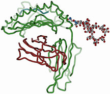
A Picture is Worth a Thousand Words
This technical innovation made it possible to obtain large
amounts of soluble protein molecules, enabling Teyton to grow
crystals and to determine the crystal structure of mouse MHC-II.
Teyton obtained structural data for two different mouse MHC
molecules: the mouse MHC-II I-Ag7, which is expressed
in the mouse model of Type 1 diabetes; and the mouse MHC-II
I-Ad, which is not linked to diabetes.
"We had hypothesized that there were unusual structural
features of I-Ag7 that accounted for its role in
autoimmunity," Teyton said. "However, after solving the structure,
we saw that the I-Ag7 is a normal class II MHC
molecule in its structural features and in the way that it
binds peptides."
Thus, many questions remain to be answered in the quest to
understand the mechanisms of diabetic autoimmunity, such as:
Is diabetes caused by a peptide-specific mechanism? The specific
antigen recognized by auto-reactive T-cells has yet to be
identified. Teyton's lab is part of a multi-center National
Institutes of Health (NIH) project that is working on this
question.
A second possibility is that the MHC-II molecule itself somehow
leads to abnormal T-cell selection. Although there are no
obvious structural differences between the MHC-II molecules
that are linked to Type 1 diabetes and those that are not,
there are potentially significant sequence differences between
them. This sequence difference results in a net negative charge
on the peptide-binding portion of the MHC-II not linked to
autoimmune diabetes, while a neutral charge in this portion
of the molecule is associated with disease susceptibility.
This difference in charge could confer selectivity for peptides
that cause diabetes, or could play a role in abnormal T-cell
selection or activation.
To further explore this possibility, Teyton is conducting
structural studies of the interaction of the diabetes-associated
MHC-II and the T-cell receptor, through the use of interference
microscopy, a new technique that allows the visualization
of single molecules at the cell surface. This enables Teyton
to examine the interactions between reconstituted T-cell receptors
and MHC-II molecules—and to detect any differences in
this interaction that are dependent on the type of MHC-II
expressed. Ultimately, this will lead to a better understanding
of the mechanisms of T-cell activation and the abnormalities
associated with autoimmune responses.
Says Teyton, "If we can detect the structural basis for a
problem, we will have a basis for specific clinical intervention.
The fundamental question remains: How are T-cells activated,
and can we specifically prevent the activation of particular
subsets?"
Teyton states his goals with intensity, recalling the frustration
of being a clinician who could not explain why his patients
were suffering. But Teyton's commitment to figuring out how
things work, combined with the advanced technological resources
at his disposal, may someday help to alleviate that frustration
for future physicians and patients.
1 | 2 |

|