Dramatic Footage of Immune System at Work Caught on Tape
By Jason Socrates
Bardi
Using a new technique that allows scientists to see the
internal machinery of a living cell, a team of researchers
at The Scripps Research Institute (TSRI) addressed one of
the most fundamental issues in immune research: the early
events in the immune system's recognition of foreign invaders,
such as bacteria and viruses, in the body.
In the latest issue of the journal Immunity, a team
led by TSRI Associate Professor of Immunology Nicholas R.J.
Gascoigne and Senior Research Associate Tomasz Zal used fluorescence
resonance energy transfer (FRET) to look at the close interaction
of immune molecules that recognize foreign antigens, which
are small molecule markers that are components of the pathogens.
Specifically, the researchers focused on the main receptor
on the surface of mature T cells, called the T cell Receptor,
and one important T cell surface "coreceptor" molecule, CD4.
In particular, Zal and Gascoigne were interested in demonstrating
vividly through FRET how other "antagonist" molecules in the
bloodstream that can bind to the T cell receptor can block
the interaction of the CD4 with the antigen, inhibit the signaling
cascade that leads to T cell activation, and reduce the effectiveness
of an immune response.
"We can look at positions of [CD4 and T cell receptor] proteins
and whether or not they are interacting," says Gascoigne.
"That allows us to see whether or not you are getting T
cell activation by a particular ligand—the very earliest
events in T cell recognition," he adds.
Recognition Key to Immune Response
The immune system long ago evolved ways to recognize pathogenic
invaders through their antigens. For instance, these antigens,
or fragments of the pathogens, may come from pathogenic proteins
that have been taken up and processed into small peptides
a few amino acids long, which are then taken up by specialized
antigen-presenting cells (APC). The APCs "present" the antigens
on their surfaces by displaying them in molecular complexes
with the so-called major histocompatibility complex (MHC)
proteins.
When a pathogen invades the immune system, APCs alert T
cells by displaying the pathogenic antigens. When specific
T cells see the antigen in the MHC, they generate a systemic
immune response designed to lead to the destruction of the
pathogen, starting with a cascade of internal activation events.
The first event in this cascade is the positive recognition
of the MHC and antigen peptide by the T cell receptor and
coreceptors. The coreceptor is crucial for this recognition
because it stabilizes the binding of the T cell receptor to
the MHC.
Once that positive recognition occurs, the T cells become
activated as killer and helper T cells, aggressively destroying
infected cells, stimulating an inflammatory response in infected
tissue, and producing chemicals that induce other cells to
make and release soluble antibodies that target the pathogen
in the bloodstream. Such immune reaction regularly keeps us
alive as we go through life in constant contact with the bacteria,
viruses, and infectious microbes of the world.
Significantly, the immune system has also evolved caution
about activating its T cells. Excessive or inappropriate immune
responses can be lethal to an organism, and so the cells of
the immune system are highly discriminating in their ability
to recognize foreign antigen and only foreign antigen. T cells
can tell the difference between foreign peptide antigen and
a "self" peptide that only differ by a single amino acid.
That one amino acid makes all the difference.
"The immune system can tell the difference," says Gascoigne,
"and it makes a totally different response."
However, the immune system can also be tricked into missing
the foreign peptide when other molecules—antagonists—block
the binding of the coreceptor to the MHC. Without this crucial
step, the T cell will not become activated even if the T cell
receptor sees the foreign antigen in the MHC.
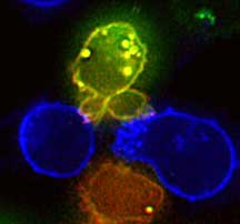
In Gascoigne and Zal's study, they use FRET to look at the
recognition of MHC by the T cell receptor and the coreceptor
CD4. They are able to see the interaction of MHC/CD4/T cell
receptor live on the screen, and find that they can block
this critical early event in immune recognition by adding
antagonists.
Fluorescence Resonance Energy Transfer
Using FRET, scientists can now look at protein–protein
interactions anywhere in a living cell in real time. FRET
works on the same basis of traditional fluorescence microscopy,
in which fluorophores—small molecules like green fluorescent
protein (GFP) that absorb and reemit photons of a particular
wavelength—are attached to proteins in the cell. One
can then illuminate the cells with a monochromatic light source
and train a microscope camera to capture the reemitted photons.
In FRET, two different fluorescent molecules are used. Under
the microscope, these two will have different emission wavelengths
and therefore different colors, cyan and yellow, for instance.
However, the emission wavelength of the cyan overlaps with
the excitation of the yellow, and so when the two molecules
are very close together, within 10 nanometers or so (a millionth
of a centimeter), the cyan molecule will donate its energy
to the yellow molecule, and yellow instead of cyan fluorescence
will result.
The new color indicates that the molecules to which the
cyan and yellow fluorophors are attached are interacting.
In the case of the Gascoigne lab's work, the CD4 molecules
had yellow fluorescent protein attached, and part of the T
cell receptor complex had a cyan fluorescent protein attached.
When the CD4 and the T cell receptor are working properly
and both recognizing the MHC, their two fluorescent proteins
are close enough to interact, which is visible as reduced
cyan fluorescence and increased yellow fluorescence upon exciting
the cyan fluorophore under the microscope. And when T cell
receptor antagonists are mixed in, there is no yellow fluorescence
from the activation of the cyan protein, which would indicate
that the fluorescent proteins—and therefore the CD4 molecules
and the T cell receptors—are not interacting.
The research article "Inhibition of T-cell receptor-coreceptor
interactions by antagonist ligands visualized by live FRET
imaging of the T-hybridoma immunological synapse" is authored
by Tomasz Zal, M. Anna Zal, and Nicholas R.J. Gascoigne and
appears in the April 17, 2002 issue of Immunity.
The research was funded by the National Institutes of Health
and the Human Frontier Science Program Organization.

|