
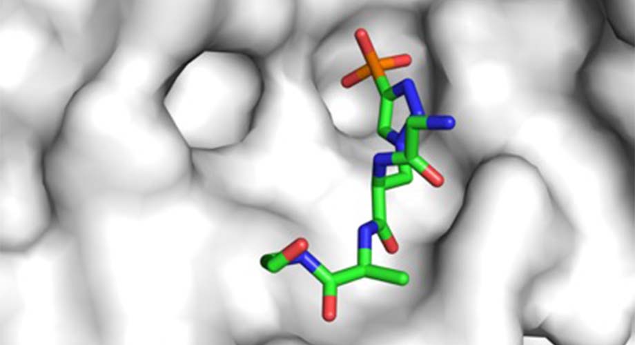
A new study shows how a specially engineered antibody (white, in background) is able to recognize different forms of phosphohistidines, molecules that are involved in the development of cancer and other conditions. (Image courtesy of the Wilson lab, Scripps Research)
Important research antibodies get their close-up
Structural biologists have created the first-ever atomic-scale maps of antibodies bound to elusive molecules that play a role in a range of human diseases.
February 23, 2021
LA JOLLA, CA—In a feat that’s been years in the making, collaborating teams from Scripps Research and the Salk Institute have revealed the atomic-scale details of how a special set of antibodies can selectively bind to elusive molecules called phosphohistidines, which are thought to play a key role in certain forms of cancer and potentially other diseases.
The discovery should enhance the use of these antibodies as powerful scientific tools for studying phosphohistidines and could help in the design of even better research antibodies. Through an enhanced understanding of phosphohistidines, scientists also can forge new medical discoveries that lead to treatments.
The study appears in Proceedings of the National Academy of Sciences.
“Through our structural studies, we were able to gain key insights into how these antibodies recognize phosphohistidines with high specificity, and thus how they can be useful as research tools,” says Robyn Stanfield, PhD, an investigator in Scripps Research’s Department of Integrative Structural and Computational Biology.
Stanfield is part of the research group led by Ian Wilson, DPhil, Hansen Professor of Structural Biology and Chair of the Department of Integrative Structural and Computational Biology at Scripps Research. She worked in concert with the Salk research team that, in 2015, succeeded in developing the set of five antibodies as a toolkit for studying phosphohistidines in the lab.
An enigmatic protein modification
A phosphohistidine is a histidine amino-acid—one of the building-blocks for proteins—with which a cluster of atoms called a phosphoryl group has reacted to form a tight bond. Cells use such phosphoryl attachments as switches to control proteins’ activities, and those that result in phosphohistidines are thought to be essential activities in many key processes in cells.
But phosphohistidines have been extraordinarily tricky to study because the phosphoryl group can attach to a histidine at either of two places, making one of two relatively unstable phosphohistidine forms, or “isomers.” Until recently, scientists have made relatively little progress in understanding them. The antibodies developed by the Salk team represent a major advance because they can selectively recognize one or the other phosphohistidine isomer, allowing each to be isolated and studied far more easily than before.
Seeing unprecedented detail
For the new study, the Salk researchers—led by Tony Hunter, PhD, American Cancer Society Professor at Salk—approached Wilson’s team, one of the world’s leading research groups for structural studies of antibody-protein complexes. Scripps Research’s Stanfield performed the structural imaging and analysis work in close collaboration with Rajasree Kalagiri, PhD, a postdoctoral researcher in the Hunter lab. The collaborating scientists used X-ray crystallography to map the atomic structures of the different isomers bound by the antibodies.
“We are fascinated with how antibodies recognize antigens and, in this case, how they are able to discriminate among small but biologically important differences in the two phosphohistidine isomers,” Wilson says. “So, we were delighted to take on the challenge of helping address this question when Dr. Hunter approached us.”
The work revealed precisely where the antibodies bind, how tightly, and how they are able to distinguish the two isomers—essentially showing how these antibodies can be useful for researching the many roles of histidine phosphorylation in health and disease.
“This fruitful collaboration with our close neighbors at Scripps Research has revealed important structural details of these antibodies,” Hunter says. “We can now use these insights to engineer the antibodies to be even more useful for studying the role of histidine phosphorylation of proteins in normal cells and in disease states.”
“Structural basis for differential recognition of phosphohistidine-containing peptides by 1-pHis and 3-pHis monoclonal antibodies” was authored by Rajasree Kalagiri, Robyn Stanfield, Jill Meisenhelder, James La Clair, Stephen Fuhs, Ian Wilson and Tony Hunter.
Funding was provided by the National Institutes of Health (5 R01 CA082683, 5 RO1 CA194584, and 1 R35 CA242443), the Leona M. and Harry B Helmsley Charitable Trust grant (2012-PG-MED002) and the Skaggs Institute for Chemical Biology at The Scripps Research Institute.
For more information, contact press@scripps.edu