
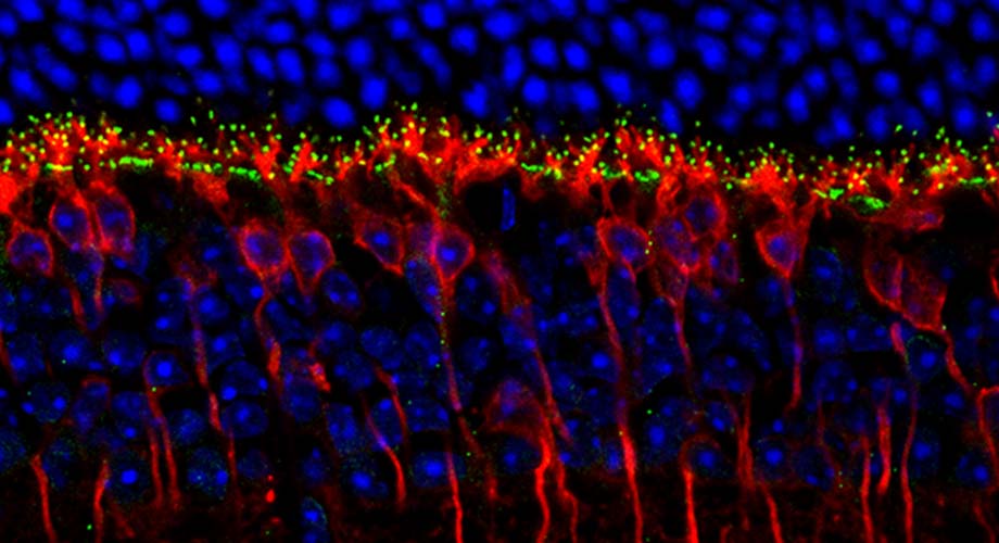
To enable vision, photoreceptors in the retina make synaptic contacts (green) with their partners called “ON bipolar neurons” (red) and transmit information to the brain. Cellular nuclei shown in blue. (Microscopy image of mouse retina courtesy of Martemyanov lab.)
Wiring the eyes to the brain for color vision
Color-discriminating cone cells in the retina use a pair of "adhesion" proteins to connect to the brain.
September 02, 2020
JUPITER, FL—Cone cells in the retina, which are meant to work in daylight and enable color vision, normally use a specific set of proteins during development to connect to other nerve cells in the retina and the brain, according to a study led by neuroscientists at Scripps Research.
The finding, reported in the Proceedings of the National Academy of Sciences, solves the mystery of how cones form their distinctive brain connections, giving humans and other animals their sophisticated and powerful vision sense. The basic neuroscience discovery may aid future efforts to boost vision lost due to retina degeneration, which is common in the elderly, and may potentially help connect lab-grown light-sensing prosthetics that cure blindness.
Solving a connection quandary
Our eyes are equipped with two kinds of light sensors known as photoreceptors: the rods that we use for seeing under very dim lighting and cones that we use ubiquitously throughout the day to see things in color.
Detecting the light, however, is only the first step for us to be able to see. Photoreceptors then need to be able to transmit their signals to the brain. They do so by establishing highly selective contacts with other neural cells in the retina that ultimately pass the information to the brain, much like plugging an electrical device into a socket.
But what makes the plug match the socket so that rod and cones can connect?
The research team, led by principal investigator Kirill Martemyanov, PhD, professor and chair of the Department of Neuroscience at Scripps Research’s Florida campus, discovered that a pair of cell adhesion molecules called ELFN1 and ELFN2 does the trick.
“It has been a thrilling scientific detective story,” Martemyanov says.
Back in 2015, his team discovered that a protein called ELFN1 enables rod cells to hook up to nearby nerve cells in the retina. A rod cell is a long, slender cell with photosensitive molecules at one end for detecting light photons, and a stalk-like projection at the other end for sending their signals brainward.
Martemyanov and his team found that rod cells, as they develop in the retina, produce ELFN1 and secrete it from their output ends—and that this induces nearby nerve cells called bipolar cells, which are connected to the brain via the optic nerve, to form connections, or synapses, with them. Once these synapses are formed, the rod cells can start sending their light-sensing signals to the vision-processing regions of the brain. The scientists found that mice engineered to lack the gene for ELFN1 were essentially “night-blind,” lacking rod-based vision but retaining cone-based vision.
That clarified how rod cells work. But what about cone cells?
“We found in that study that cone cells normally don’t express ELFN1,” Martemyanov says, “so we started looking for a protein like ELFN1 that is produced in cone cells and has a similar role in inducing connections between these cells and cone-specific bipolar cells.”
A clearer view of the vision system
The process of finding this ELFN1-like cone protein required studies using animals whose vision sense is dominated by cone cells, not rod cells—whereas mice and even humans use mostly rod cells. Martemyanov and his team therefore studied cone-dominant ground squirrels and tree shrews to identify the protein that does for cones what ELFN1 does for rods. The protein was already considered such a close relative of ELFN1 that it had been named ELFN2.
But the hunt was not over, because when Martemyanov and his team engineered “knockout” mice that lack the gene for ELFN2, the retinas of the mice developed normally, with apparently functional cone cells.
Eventually, the researchers were able to determine that ELFN1, though it normally isn’t produced detectably in cones, is produced in cones to perform ELFN2’s wiring function if ELFN2 is missing.
It’s “a classic example of resilience in an important biological system,” Martemyanov says.
With the new discovery, he says, we finally know how two types of primary photoreceptor cells, which we rely on for all of our vision sense, are sending their information to the brain. And importantly, this fundamental knowledge may one day translate into novel treatments for vision loss.
“Interplay between cell adhesion molecules governs synaptic wiring of cone photoreceptors” was authored by Yan Cao, Yuchen Wang, Henry Dunn, Cesare Orlandi and Kirill Martemyanov, of Scripps Research; Nicole Shultz, Naomi Kamasawa, and David Fitzpatrick, of Max Planck Florida Institute; Wei Li of the National Eye Institute; Christina Zeitz of Sorbonne Université; and William Hauswirth of the College of Medicine at the University of Florida at Gainesville.
The study was funded by the National Institutes of Health (EY018139 and EY028033), the French Muscular Dystrophy Association, Retina France and the Agence Nationale de la Recherche.
For more information, contact press@scripps.edu