(page 2 of 2)
This work started a few years ago after Ginsberg's group
discovered a salt bridge between the alpha and beta tails
of the integrin. Salt bridges are favorable interactions formed
by two oppositely-charged ionized groups within a protein,
and Ginsberg knew that this salt bridge probably had a stabilizing
effect on the interface between the two subunits—locking
them in place, so to speak.
Ginsberg demonstrated that if he mutated the amino acid
residues that formed this salt bridge disrupting these contacts,
the integrins became activated.
This led him to speculate that under normal conditions,
some sort of association between the inner tails of the alpha
and beta subunits of the integrin held the protein in an inactive
position.
"Whatever was activating [the integrins]," says Ginsberg,
"was doing it by pulling the tails apart."
Around the same time, Calderwood arrived at TSRI as a postdoctoral
fellow in the Ginsberg lab. Calderwood and Ginsberg began
asking what cellular proteins might be disrupting the two
tails of the integrin subunits, and they soon focused on talin.
Talin is a large intracellular protein more than 2,000 amino
acids long and a major cytoskeleton protein on the inside
of cells. Most of the talin protein binds to actin—the
filamentous cellular protein that makes up the cytoskeleton
and gives a cell its shape.
But Calderwood and Ginsberg discovered a small domain on
the amino-terminus end of talin that binds to the beta-subunit
tail of the integrin.
A Fruitful Collaboration
This week's report in Science shows that talin, indeed,
is essential to integrin activation. The report is the result
of a fruitful collaboration between Calderwood, Ginsberg and
several other scientists at TSRI and The Burnham Institute.
TSRI Professor Sanford Shattil and his former TSRI postdoctoral
fellow Seiji Tadakoro contributed their expertise with a technique
called RNA interference.
RNA interference involves delivering small, 20- to 30-base
pieces of double-stranded RNA into a cell. Once inside the
cell, these short sequences anneal to complementary regions
of cellular RNA and trigger an intracellular response that
specifically destroys the target RNA. The technique allows
scientists to selectively shut off normal cellular genes and
permits them to study the impact of the absence of the corresponding
gene products on cellular function.
The team used RNA interference to remove the talin from
a type of cell called a megakaryocyte, a precursor of platelets,
which TSRI postdoctoral fellow Koji Eto derived from embryonic
stem cells. These megakaryocytes have the same machinery as
platelets and respond to certain stimuli the same way that
platelets do.
One of these stimuli is the chemical adenosine 5' diphosphate
(ADP). When platelets are exposed to ADP, they become activated
and the integrins on their surface switch from low to high
affinity. The same is true of the megakaryocytes.
However, Calderwood and his colleagues showed that when
the talin was removed from the megakaryocytes by RNA interference,
the ADP no longer worked.
"It could not activate the integrins," says Calderwood,
adding that they were able to rescue the activation by adding
talin back into the cells from which it had been removed.
"This is a great example of a [scientific] collaboration,"
says Shattil. "It provided the critical evidence that talin
was required for integrin activation."
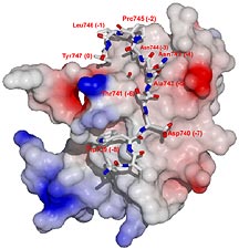
The TSRI scientists also collaborated with Robert C. Liddington
and Jose M. de Pereda of The Burnham Institute, with whom
they had previously solved the crystal structure of talin
bound to the cytoplasmic domain of integrin. This structure
enabled Liddington and de Pereda to suggest places to mutate
the talin and the beta subunit of the integrin to selectively
disrupt the interaction between the two proteins.
"When [TSRI Research Assistant] Vera Tai introduced those
mutations into full-length integrins, those integrins are
inactive," says Calderwood. In the paper, the team also points
out that overexpressing talin normally activates integrins.
Overexpressing the mutant form of talin has no effect.
Further Questions
The importance of this discovery is enhanced by the fact
that talin binds to almost all of the various tails of the
beta subunits of integrins (eight of which are known).
The next step for Calderwood, Ginsberg, Shattil and their
colleagues is to ask how the cell controls talin binding.
Figuring out these mechanisms is particularly interesting
from a therapeutic point of view, since integrins are involved
in such major killers as heart disease and cancer. Because
talin binding is the final step in integrin activation, it
might be a good target for keeping the integrins from becoming
active.
"It's theoretically possible to perturb this interaction
pharmaceutically," says Calderwood.
1 | 2 |

|