| |
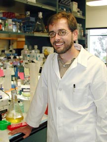
The Tail End of Integrin ActivationThere is no shortage of information on the Internet about integrins. A recent Google search for the word "integrin," in fact, turned up 263,000 web pages devoted to the structure, chemistry, and biology of this important family of cell-surface proteins, which are involved in everything from early embryonic development to the development of heart diseases and cancer later in life. There was even one site that boasted an integrin chat room. Such an ocean of preexisting information begs the question, is there anything more to say? In this case, the answer is an emphatic yes. Written by a team of scientists from The Scripps Research Institute (TSRI) and its neighboring La Jolla institution, The Burnham Institute, a paper appearing in this week's issue of the journal Science describes a crucial final step in the process of integrin activation—the binding of a protein called talin. "Talin is required for the activation process," says TSRI Assistant Professor David Calderwood, who led the study. "This interaction is the last step." The study is interesting because understanding the way in which integrins are activated is crucial to understanding their function in all the physiological processes in which integrins are involved. Integrins and PlateletsIntegrins are large binary protein complexes made up of two different types of polypeptide chains (called the alpha and beta subunits) that come together to form a "heterodimer" that is expressed on the surface of a cell. They are somewhat top-heavy. A huge portion of the protein is extracellular and sticks out on the outside of the cell, and just a tiny tail of a few dozen amino acids protrudes through the membrane on the inside of the cell. The large extracellular portions are the domains that bind to molecules on the outside of the cells and mediate the interactions of the cell with other cells. If tissues were trains and cells were the boxcars, then integrins would be the hooks that hold the boxcars together. They hold cells together and keep them bound to one another and to the extracellular matrix maintaining the integrity of tissues in mammals and other multicellular organisms. They are also important in early development for the formation of distinct tissues. But integrins do more than just hold cells together. They are also crucial mediators of a host of other normal and abnormal biological processes. They are important for inflammation; they are essential for platelet aggregation after vascular injury; and they are involved in cell motility. As such, they are involved in diseases where the normal mechanisms of platelet aggregation go awry—as in heart attacks and strokes—and are implicated in cancer metastasis. Not surprisingly, scientists have for years been interested in what integrins do, how they are involved in conditions like cancer, heart attacks, and stroke, and whether the mechanisms of integrin activation could be modulated to improve the prognosis of patients. For instance, one of the molecules to which integrins bind is fibrinogen, a circulating dimeric protein that is present in large amounts in the blood and can bind integrins at both ends. This interaction is essential for mediating the aggregation of platelets—those flat, molecule-filled cytoplasmic disks in the blood. Platelets are covered with integrins (typically 80,000 are on the surface of any given platelet). But the integrins need to be activated to bind fibrinogen. When they are not active, the platelets flow in the blood without sticking to each other or to blood vessel walls. An injury will cause the integrins on the surface of platelets to become activated. The activated integrins then bind to fibrinogen, which then bind to other activated integrins on other platelets, cross-linking many platelets into a massive thrombus. The body tightly controls this cascading reaction. Not enough thrombus formation could lead to massive blood loss, and too much could lead to a lethal, occlusive thrombus, causing a heart attack or stroke. Understanding how integrins are activated, then, is a crucial question for scientists. Calderwood and his Department of Cell Biology colleagues, TSRI Professors Mark Ginsberg and Sanford Shattil investigated this topic thanks to support from the Program in Hemostasis and Thrombosis at the National Heart, Lung, and Blood Institute, one of the National Institutes of Health, and from the American Heart Association. The Vital Step in Integrin ActivationHow exactly the activation of integrins is controlled by the body has been an open question for several years, but in the last decade more and more evidence has pointed to the importance of the tiny tails of the integrins inside the cells. How these small cytoplasmic domains activate integrins has been studied for some time. In fact, says Ginsberg, many—perhaps thousands—of scientific papers published on various steps in the pathway of integrin activation and molecules that perturb these steps. What has not been known, until now, is the final step in this activation process. The talin protein turns out to be key. Though the mechanism is not completely clear, Calderwood, Ginsberg, and their colleagues have evidence that shows when talin binds to the beta subunit of integrin, it causes a conformational change in the integrin, which is propagated across the membrane, changing the structures of the integrin domains on the outside. "This is the vital step," says Calderwood. "Talin binds to the cytoplasmic tail, and that passes a signal that changes the large extracellular domain." This work started a few years ago after Ginsberg's group discovered a salt bridge between the alpha and beta tails of the integrin. Salt bridges are favorable interactions formed by two oppositely-charged ionized groups within a protein, and Ginsberg knew that this salt bridge probably had a stabilizing effect on the interface between the two subunits—locking them in place, so to speak. Ginsberg demonstrated that if he mutated the amino acid residues that formed this salt bridge disrupting these contacts, the integrins became activated. This led him to speculate that under normal conditions, some sort of association between the inner tails of the alpha and beta subunits of the integrin held the protein in an inactive position. "Whatever was activating [the integrins]," says Ginsberg, "was doing it by pulling the tails apart." Around the same time, Calderwood arrived at TSRI as a postdoctoral fellow in the Ginsberg lab. Calderwood and Ginsberg began asking what cellular proteins might be disrupting the two tails of the integrin subunits, and they soon focused on talin. Talin is a large intracellular protein more than 2,000 amino acids long and a major cytoskeleton protein on the inside of cells. Most of the talin protein binds to actin—the filamentous cellular protein that makes up the cytoskeleton and gives a cell its shape. But Calderwood and Ginsberg discovered a small domain on the amino-terminus end of talin that binds to the beta-subunit tail of the integrin. A Fruitful CollaborationThis week's report in Science shows that talin, indeed, is essential to integrin activation. The report is the result of a fruitful collaboration between Calderwood, Ginsberg and several other scientists at TSRI and The Burnham Institute. TSRI Professor Sanford Shattil and his former TSRI postdoctoral fellow Seiji Tadakoro contributed their expertise with a technique called RNA interference. RNA interference involves delivering small, 20- to 30-base pieces of double-stranded RNA into a cell. Once inside the cell, these short sequences anneal to complementary regions of cellular RNA and trigger an intracellular response that specifically destroys the target RNA. The technique allows scientists to selectively shut off normal cellular genes and permits them to study the impact of the absence of the corresponding gene products on cellular function. The team used RNA interference to remove the talin from a type of cell called a megakaryocyte, a precursor of platelets, which TSRI postdoctoral fellow Koji Eto derived from embryonic stem cells. These megakaryocytes have the same machinery as platelets and respond to certain stimuli the same way that platelets do. One of these stimuli is the chemical adenosine 5' diphosphate (ADP). When platelets are exposed to ADP, they become activated and the integrins on their surface switch from low to high affinity. The same is true of the megakaryocytes. However, Calderwood and his colleagues showed that when the talin was removed from the megakaryocytes by RNA interference, the ADP no longer worked. "It could not activate the integrins," says Calderwood, adding that they were able to rescue the activation by adding talin back into the cells from which it had been removed. "This is a great example of a [scientific] collaboration," says Shattil. "It provided the critical evidence that talin was required for integrin activation." The TSRI scientists also collaborated with Robert C. Liddington and Jose M. de Pereda of The Burnham Institute, with whom they had previously solved the crystal structure of talin bound to the cytoplasmic domain of integrin. This structure enabled Liddington and de Pereda to suggest places to mutate the talin and the beta subunit of the integrin to selectively disrupt the interaction between the two proteins. "When [TSRI Research Assistant] Vera Tai introduced those mutations into full-length integrins, those integrins are inactive," says Calderwood. In the paper, the team also points out that overexpressing talin normally activates integrins. Overexpressing the mutant form of talin has no effect. Further QuestionsThe importance of this discovery is enhanced by the fact that talin binds to almost all of the various tails of the beta subunits of integrins (eight of which are known). The next step for Calderwood, Ginsberg, Shattil and their colleagues is to ask how the cell controls talin binding. Figuring out these mechanisms is particularly interesting from a therapeutic point of view, since integrins are involved in such major killers as heart disease and cancer. Because talin binding is the final step in integrin activation, it might be a good target for keeping the integrins from becoming active. "It's theoretically possible to perturb this interaction pharmaceutically," says Calderwood.
|
|
|