Targeting Breast Cancer Metastasis
By Jason Socrates Bardi
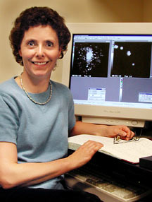
Associate Professor Brunhilde Felding-Habermann recently
sat in her office going over some slides she was preparing
about her work on metastatic melanoma and breast cancer cells.
She was getting ready to give a talk this week at the 2003
annual retreat of the Department of Molecular and Experimental
Medicine (MEM) at The Scripps Research Institute (TSRI).
"I have titled this talk "Targeting Breast Cancer Metastasis,"
says Felding-Habermann.
She adds that breast cancer is still a pressing public health
concern because it has a high propensity to metastasize and
when it does, it can be deadly. "Too many patients still die
from metastatic breast cancer," she says.
A Dangerous Phenomenon
Metastasis is a dangerous phenomenon in which cancer cells
separate from a tumor mass, move through the bloodstream,
anchor down in a distant tissue or organ, and begin a new
cancer that might compromise the function of that organ. While
surgeons can remove cancerous tissue, such procedures are
greatly complicated if a tumor establishes new tumors in other
organs. Although science and medicine have made tremendous
strides in early detection and successful treatment, breast
cancer and melanoma still claim tens of thousands of lives
a year—usually the end result of metastasis.
The Centers for Disease Control and Prevention (CDC) reports
that by the end of 2003, 211,300 U.S. women will be diagnosed
with new cases of invasive breast cancer and that an estimated
39,800 women will die of the disease this year.
Similarly, melanoma, the deadliest type of skin cancer,
claims many lives. According to the CDC, some 54,000 patients
in the United States will be diagnosed with new cases of melanoma
in 2003 and 7,700 Americans will die of the disease.
What leads cells to metastasize? What determines where the
metastatic cancer cells go? Why do breast cancers typically
metastasize and form new tumors in the brain, lungs, liver,
and bones specifically? What is it about the vascular cells
in these tissues that interacts with the breast cancer cells
and allows them to invade, forming a new tumor?
Answering these questions is where basic biomedical scientists
like Felding-Habermann and her Scripps colleagues come in.
"Disseminating tumor cells suddenly find themselves having
to demonstrate capabilities that they did not need to have
in the primary tumor," says Felding-Habermann. "What we are
trying to find out is what determines whether or not breast
cancer or melanoma cells will metastasize."
Readying the Invasion
Such specific questions are the key to understanding cancer
because cancer is a complicated disease. It can be caused
by subtle mutations within normal cells. After certain mutations
occur, a cancer cell grows resistant to normal programmed
cell death, dividing out of control, over and over, and forming
a solid tumor. Also common to tumor cells are mutations that
lead them to metastasize.
The word "metastasis" comes from the Latin construction
meaning to change position, and metastasis depends
on the cancerous cells acquiring two separate abilities—increased
motility and invasiveness.
Motility, the ability of a cell to get up and go, is an
obvious requirement for metastasis. Perhaps not so obvious
is the need for cancerous cells to invade tissue, to burrow
in and set up shop somewhere else.
These "phenotypic" abilities do not arise out of thin air.
Cancerous cells acquire them in part through mutations to
their DNA that result in changes of expression of one or more
genes. Some mutations turn on or increase the activity of
certain key genes, increasing the expression of metalloproteinases
for instance; others downregulate them, shutting off production
of receptor proteins. Often as these expression levels change,
so do the levels of other, related genes.
Changes in gene expression that seem to control metastasis
in human breast cancer affect an adhesion receptor called
integrin avb3.
Integrins are large protein complexes made up of two different
types of polypeptide chains (called the alpha and beta subunits)
that come together to form a "heterodimer" that is expressed
on the surface of a cell.
They are somewhat top-heavy. A huge portion of the protein
is extracellular and sticks out at the surface of the cell,
and just a tiny tail of a few dozen amino acids protrudes
through the membrane on the inside of the cell.
The large extracellular portions are the domains that bind
to molecules on the outside of the cells and mediate the interactions
of the cell with surrounding matrix proteins and with other
cells.
If tissues were trains and cells were the boxcars, then
integrins would be the hooks that hold the boxcars together
and keep them on their tracks. This helps to maintain the
integrity of tissues in mammals and other multicellular organisms.
They are also important in early development for the formation
of distinct tissues.
But integrins do more than just hold cells together. They
are also crucial mediators of a host of other normal and abnormal
biological processes. They are important for inflammation;
they are essential for platelet aggregation after vascular
injury; and they are involved in cell motility. As such, they
are involved in diseases where the normal mechanisms of platelet
aggregation go awry—as in heart attacks and strokes—and
they are implicated in cancer metastasis.
Capturing the Arrest of Tumor Cells
One reason why integrins are important for metastasis is
that they help the cancer cells attach within the vasculature
of new tissues.
When a cancer cell is in the circulation, it may have difficulty
locking onto new tissues because of the interference of blood
cells, plasma proteins, and the shear force produced by blood
flow. Cancer cells need something like a hook to anchor themselves
to a particular spot under these dynamic flow conditions,
and integrins can be such hooks.
Felding-Habermann uses a video microscopy system with a
flow chamber, established by MEM Professor Zaverio M. Ruggeri,
to study how tumor cells arrest in flowing blood. The surface
in the flow chamber is engineered to mimic the surface of
a blood vessel—with a layer of endothelial cells on top
of collagen and other matrix components. When she began these
experiments, her question was what would happen when she added
metastatic breast cancer cells to blood and "perfused" this
mixture over components of the vessel wall.
Her videos show the results: the movement of blood across
the screen. Colored with a green fluorophor are platelets,
those tiny, molecule filled cells in the blood that are necessary
for clotting. Also moving across the surface are cancer cells,
which are colored with a red fluorophor. When highly aggressive
tumor cells are mixed into the blood sample, they get stuck
to the surface. Non-metastatic cells just move on with the
flow. Figuring out why was the challenge. Felding-Habermann
noticed that the platelets, which form tiny clots or thrombi,
help the aggressive cancer cells to arrest.
"You see all these thrombi?" she asks, pointing at the green
blotches on the screen. "Almost all of them have tumor cells
stuck to them or embedded in them. You notice that when I
switch to the microscope filter that detects only the red
signal from the tumor cells."
The Link Between Integrins and Cancer
Breast cancer cells express a particular integrin, called
avb3,
that is similar to an integrin expressed on the surface of
platelets. Normal breast epithelium does not express this
integrin, but cancer cells do.
Integrin avb3
seems to be needed for the cancer cells' arrest.
The cancer cell integrin has the ability to support interactions
between the tumor cells and platelets by binding plasma proteins.
Once bound, these proteins can form molecular bridges between
the tumor cell integrins and platelet integrins. Felding-Habermann
noticed that tumor cell integrin avb3
supports this interaction while blood is flowing - but only
if the receptor is activated, as on aggressive, metastatic
cells.
This way, tumor cells with activated integrins on their
surfaces can cross-link when they hit a thrombus of platelets,
even if they are tiny, and they can readily attach to the
tissue at that location.
"What you get is a cohesion of tumor cells and platelets,"
says Felding-Habermann.
The tumor cells with active integrins on their surface can
also fish out platelets from the bloodstream. Once the tumor
cells have platelets on their surface, they become very sticky.
This is shown on another one of her videos. Green and red
globs pass together over the surface, getting stuck from time
to time as they flow. These globs can form microaggregates
as another strategy for arresting, she says. The microaggregates
can get stuck in narrow capillaries where the tumor cell can
then establish a new colony.
To demonstrate that these integrins were necessary for tumor
cell arrest, Felding-Habermann and her colleagues selected
a cancer cell line without the beta subunit of the integrin.
She discovered that those cells cannot arrest on the surface,
but if the beta subunit was added back in, the ability of
the cancer cells to arrest was restored.
Similarly, Felding-Habermann says that she has compared
normal blood to blood without platelets. The platelets in
normal blood immediately begin to clump under the microscope,
and the cancer cells soon attach to these clumps. However,
she found that when one removes the platelets from the bloodstream,
the breast cancer cells cannot attach.
To test the clinical relevance of these findings, Felding-Habermann
collaborates with physician Alan Saven, director of the Scripps
Cancer Center and Head, division of Hematology/Oncology, Scripps
Clinic. Saven provides the clinical insight and connects her
with clinicians who help her collect blood samples and tissue
specimens from patients with metastatic breast cancer. Felding-Habermann
has fished out metastatic tumor cells from patient blood and
demonstrated that they do indeed interact with platelets by
using integrin avb3.
Her perfusion experiments proved that all of the isolated
metastatic cells carry avb3
in its activated form.
1 | 2 |

|