Microscopy of Live Cells in Motion
Seest
thou her locks, whose sunny glow
Half shows, half shades, her neck of snow?
———Sir
Walter Scott, Ivanhoe
By Jason Socrates Bardi
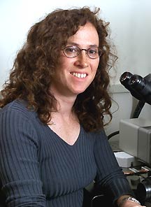
Cells are in motion on the computer monitor in the office
of Clare Waterman-Storer, who is assistant professor in the
Department of Cell Biology and the Institute for Childhood
and Neglected Diseases.
Big speckled cells—awash with activity. She is showing
a video of the cells she recorded earlier and is busy pointing
out some of their more subtle features: These are probably
actin bundles moving around organelles, here is where the
nucleus should be (off-screen above the monitor), this is
how large the cell really is (about four times larger than
the screen), and this is cell’s leading edge, she says,
pointing to a speckling mass in the lower left hand of the
screen.
Actin filaments polymerizing along the leading edge and
moving backwards to the cell center. They look like a waterfall
but act more like a treadmill, she tells me.
“I love seeing these movies,” she says.
Waterman-Storer came to The Scripps Research Institute (TSRI)
last year to start up the Laboratory for Cell Motility, an
area that has important implications for many fields.
She studies the molecules that are involved with cell motility—particularly
microtubules, actin, and all the proteins responsible for
regulating them. In particular, she is interested in the structural
and regulatory interactions between actin and microtubules—how
they touch and move each other and how they affect each other.
Her laboratory is specifically interested in how these molecules
interact during the control of motility and how those interactions
impact such diverse areas as cancer, wound healing, and early
embryonic development.
In order to address these questions, Waterman-Storer uses
a microscopy method with which she can image both the microtubules
and actin directly. She can also quantitatively assess what
happens to these structural proteins in living cells when
she makes changes to the signaling and regulatory proteins
that bind to them.
This method, called fluorescence speckle microscopy (FSM),
was developed by Waterman-Storer and her former boss E.D.
Salmon while she was doing a post doctoral fellowship is his
laboratory at the University of North Carolina at Chapel Hill
in the late 1990s.
"We discovered it by accident," she says.
Like Looking at a Brick Wall
Waterman-Storer and Salmon had been using another technique,
called fluorescence analog cytochemistry, to study actin and
microtubule motion in the cell.
In this technique, fluorophores are covalently attached
to actin or microtubule subunits and then microinjected back
in the cell. Fluorophores are simply small molecules that
absorb and reemit photons of a particular wavelength.
One can then illuminate the cells with a monochromatic light
source and train a microscope camera to capture the reemitted
photons. For instance, one can attach green florescent protein
to the tubulin subunits from which microtubules, which are
shaped somewhat like brick chimneys, are constructed.
But this technique always had problems imaging the cytoskeleton
in living cells because of the high concentration of fluorophores.
An overabundance of labeled subunits would be injected into
a cell so that the microtubules assembled with a high concentration
of these subunits. But the microtubules could never assemble
all the fluorescing subunits. This would cause too much background
florescence to see individual filaments in certain parts of
the cell.
Another, related, problem was that fluorescence analog cytochemistry
could not resolve microtubule motion because the microtubules
would be too evenly labeled with fluorescing molecules.
"It’s like looking at a brick wall from a distance,
where you cannot see the individual bricks and they are all
red," she says. "That wall could be moving in front of you,
but if you can’t resolve the individual bricks, all you
would see is a red wall, and you wouldn’t know if it
were moving or stationary."
FSM seeks to resolve the movement of the wall by painting
only certain bricks white. Then the movement of the wall could
be followed by tracking the position of the white bricks.
In fact, FSM uses about 100 times less fluorescent material
than fluorescence analog cytochemistry—only about one
tenth of one percent of the subunits are labeled—but
the few that are show how the whole wall moves.
"FSM," says Waterman-Storer, "Is basically fluorescence
analog cytochemistry with less florescence.
This may sound simple, but people had been using the technique
of fluorescence analog cytochemistry for over 20 years, and
for years they had occasionally injected too little fluorescing
material into the cells. Over and over, researchers wound
up with spotty, or speckled microtubules, and started over.
"We realized that this was something that could give us
information," says Waterman-Storer. This realization was enough
to turn what were formerly regarded as anomalous mistakes
into a new technology.
Using FSM, the growing actin or microtubule molecules appear
speckled, and the movement of these speckles stands out to
the eye, making it apparent. One can watch the growth of the
protein bundles and their retrograde flow, which is thought
to pull the cells along.
1 | 2 |

|