The General Assembly of Retroviruses
By Jason Socrates
Bardi
Under the electron microscope, different retroviruses often
look different because of the shapes of their capsids—protein
shells that surround the retroviral RNA on the inside of the
virus.
Human immunodeficiency virus (HIV) has a cone-shaped capsid,
for instance, whereas the Rous sarcoma virus capsid is shaped
more like a sphere. Some retroviruses have capsids that are
shaped like rods. Even different HIV subtypes can have distinct
appearances under the electron microscope.
But these differences among retrovirus capsids belie an
underlying similarity among them.
Even though the shapes of different capsid shells vary,
they may assemble using one common mechanism, reports a team
of scientists led by investigator Mark Yeager at The Scripps
Research Institute (TSRI) and investigator Wesley Sundquist
at the University of Utah. Yeager, Sundquist, and their colleagues
recently published a paper in the European Molecular Biology
Organization Journal that proposes a general model for
retroviral capsid assembly.
This research is significant because understanding how the
virus matures may be important for finding targets for intervening
in this process. Stopping the maturation of HIV has already
been proven as a therapeutic strategy through protease inhibitors,
which process the Gag precursor proteins. Inhibitors that
block the formation of the hexameric capsid lattices might
prove an effective complement to existing antiretrovirals.
Capsid Catch Can
Retroviruses contain a dozen or so genes, and these enable
the various stages of the virus lifecycle—from the initial
entry into a new cell to the replication and formation of
new virus particles late in the lifecycle.
When retroviruses form infectious "virions," they do so
by first expressing their RNA and protein components and then
assembling these molecular components on the inside of an
infected cell. These components will bind to the cell membrane
and assemble there, leading to the budding off of an immature
virion. Inside the immature virion are long Gag polyproteins,
which are precursors of the structural proteins nucleocapsid,
capsid, and matrix.
After budding, the virion must still "mature" into an active,
infectious particle by using a protease to chop the precursor
Gag proteins into their component pieces. Once these structural
proteins are free, they can self-assemble into the structures
that give a retrovirus its classic shape and appearance. Capsid
proteins, for instance, assemble into the protein capsid shell
that surrounds the RNA of the retrovirus and appears as a
cone in HIV and as a sphere in Rous sarcoma virus.
The capsid proteins from several different retroviruses
have now been solved, and their 3-D structures are virtually
identical. However despite these similarities, different retroviruses
appear quite different under the electron microscope. Somehow
the retroviruses achieve completely different forms from the
same ingredients—like two different chefs who both cook
with the same rice, fish, and vegetables but make, respectively,
a dish of fried rice and a dish of sushi.
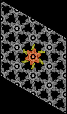
According to the new general model, capsid proteins achieve
their final form by assembling first into honeycomb-like hexameric
lattices. The Sundquist lab had previously suggested that
all retroviruses use this same lattice to produce their capsids.
The characteristic rod, cone, and spherical shapes are generated
by the insertion of pentamers of molecules within the hexameric
lattice.
When you put a pentamer into a hexameric lattice, you cause
that lattice to bend. Enough of these pentamers will close
the lattice. This situation is analogous to a soccer ball,
which relies on pentagons of leather sewn onto the sides of
hexagons to achieve a shape approximating a sphere.
In a retrovirus, a relatively even distibution of pentamers
in the hexameric lattice will generate a spherical shape.
Similarly, a rod or cone-shaped hexameric lattice can be closed
by inserting pentamers at the ends of the lattice in a defined
pattern.
Barbie Ganser, a graduate student with Wes Sundquist, visited
the Yeager lab and grew 2D crystals of capsid molecules from
the Moloney murine leukemia virus (M-MuLV), a virus that forms
a spherical capsid. They found that the packing of capsid
domains in M-MuLV conforms to a general model that had originally
been developed as a way of describing HIV virus assembly.
They propose a general model for the assembly of retrovirus
capsid molecules with the mature virion.
According to the model, the structures of the capsid protein
domains are more or less the same in the various retroviruses.
The packing of these domains into hexameric lattices is the
same as well, but flexible linker motifs between domains of
individual capsid proteins must somehow produce the different
shapes and sizes of the final products by controlling the
location of pentamers in the hexameric lattice.
To read the article, "Three-dimensional structure of the
M-MuLV CA protein on a lipid monolayer: a general model for
retroviral capsid assembly" by Barbie K. Ganser, Anchi Cheng,
Wesley I. Sundquist, and Mark Yeager, please see The
EMBO Journal Vol. 22, pp. 2886–2892, 2003.

|