|
(page 2 of 2)
By doing these experiments, they were able to go beyond
simply asking which genes are upregulated and which are downregulated
in the tumor cell. Instead, they are determining which genes
are regulated as a direct result of FAK expression.
Furthermore, Schlaepfer and his colleagues established in
vivo models in which they can effectively take away the
ability of FAK to invade tissues. They used an inhibitor of
FAK activity to selectively disrupt the invasion component
alone. The inhibitor is actually just a fragment of the FAK
gene itself that competes with endogenous FAK for binding
to integrins.
“We’re throwing a wrench into the FAK signaling
system to answer the question, if we stop its function, what
happens?” says Schlaepfer.
Interestingly, they found that stopping FAK takes away,
from tumor cells, the ability to metastasize but does not
affect their motility. This enabled them to dissociate the
role of FAK in motility versus its role in invasion. It also
led to an interesting direction for the research.
FAK in Motility and Invasion
FAK has a role to play in motility and invasion because
it is present in the projections that cells form when they
are invading new tissue. In the parlance of cell biologists,
these feet are referred to as “invadopodia” or “pseudopodia”
Podia, in Latin, means feet.
Pseudopodia are foot-like extensions that cells use for
probing an area and crawling. And within these pseudopodia,
FAK is highly expressed. Staining cells growing in culture
for phosphotyrosine, a sure sign of FAK activity, will show
hotspots at the ends of actin filaments, where the FAK signaling
is taking place.
“During invasion, these same feet squeeze between cells,”
says Mitra. “We’ve seen FAK specifically enriched
[in invading cell extensions].”
Another important cancer enzyme that is often overexpressed
in cancer cells and is localized to pseudopodia are enzymes
known as matrix metalloproteinases (MMPs).
MMPs are secreted enzymes that play a number of important
biological roles in both the early development of organ structures
and in tissue remodeling. Their physiological function is
to remodel the extracellular matrix, and because of the potential
damage that this could do to tissues, MMPs are one of the
most highly regulated enzymes in the body.
“If they weren’t regulated,” says Schlaepfer,
“our bodies would dissolve, basically.”
Unfortunately, this sophisticated regulation does not prevent
cancer cells from subverting MMPs for their own purposes—cancer
cells secrete these enzymes in order to break free of the
extracellular matrix and tissue stroma, allowing them to move.
It also allows them to dissolve barriers that come in their
way to the bloodstream or to distant tissues during metastasis.
Significantly, when FAK is upregulated in a tumor cell,
that cell will correspondingly upregulate MMP expression and
activity as well. This leads to the tantalizing possibility
that FAK is one of the signaling proteins that cancer cells
use to activate MMPs and achieve metastasis. Schlaepfer and
colleagues are testing the connections between FAK, MMPs,
and metastasis.
“If we can figure out how FAK is functioning, and if
we can get a good inhibitor then we might be able to stop
cells from metastasizing,” says Schlaepfer. “These
drugs might contain a tumor, preventing it from spreading
if it is found early enough.”
In addition to the regulation of MMPs, Schlaepfer is also
looking at the effect of FAK inhibition on certain other genes
within the cells. Looking at these “peripheral”
markers that are up- or down-regulated by FAK expression,
might be the easiest way to gauge the effectiveness of a future
FAK inhibitor in vivo and could be a useful application
for testing whether any given FAK inhibitor works in a clinical
setting.
1 | 2 |

|


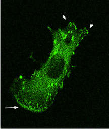 This
FAK-null tumor cell has been reconstituted with FAK and stained
to show where the protein localizes. The staining shows FAK
concentrated in areas of focal contact (top arrowheads) and
of pseudopodia formation (lower arrow).
This
FAK-null tumor cell has been reconstituted with FAK and stained
to show where the protein localizes. The staining shows FAK
concentrated in areas of focal contact (top arrowheads) and
of pseudopodia formation (lower arrow).