|
(page 2 of 2)
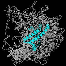
Prions Under the Magnet
One of the largest areas of study in Wüthrich's laboratory
involves prion proteins. Mis-folded prion proteins have been
suggested to cause bovine spongiform encephalopathy, or mad
cow disease, and a form of the same disease in humans, called
variant Creutzfeldt-Jakob Disease.
Prion proteins are expressed widely throughout the body
and sit anchored onto the surfaces of cells in a wide variety
of tissue, particularly on cells in neuronal tissue.
Infectious, malformed prion proteins start out with one
shape, which is innocuous, and end up with another shape,
which is observed in organisms suffering from a deadly "prion"
infection. Infectious prions from an animal with mad cow disease,
for instance, are believed to transmit the disease by initially
causing normal prion proteins in the brain of a healthy cow
to misform into the infectious form. Then these prions will
act on more normal prion proteins to produce more and more
misfolded proteins that accumulate and eventually lead to
a sponge-like build-up and brain damage.
Wüthrich concentrates on comparative studies of the
normal form of the prion protein in various species.
"[We want] to get the molecular basis of the species barrier,"
says Wüthrich. "Why are there no records of transmission
from sheep to man, but there is mounting evidence that there
is transmission from cattle to man?"
The assumption is that the more similar the prions are across
species, the easier the transmission will be, as in cows to
humans.
"Our results so far show that the healthy forms of the prion
protein between man and cattle are identical in the folded
part of the molecule," says Wüthrich. Other species,
he adds, show differences even though the global fold is maintained.
Wüthrich does not stop at cows. He has solved the structure
of the prion protein from chickens, for instance, and he is
almost done with that of the turtle.
Prions are interesting also because much of the molecule
is unstructured. The protein has a long tail that is highly
flexible and is as much at ten times longer than the diameter
of the folded part of the protein. "By studying evolutionarily
widely divergent species, we hope to possibly target some
clues as to the natural function of the prion protein, anticipating
that the active site would be preserved," says Wüthrich.
Wüthrich is also interested in making preparations
of prion protein aggregates that could be used to study the
misfolded protein and the molecular basis of the aggregation.
The needs are tantalizingly simple: a sample of isotope-labeled
prion protein in solution that form repetitious aggregates.
But the difficulty is preparing a sample that aggregates only
a little, from two to forty proteins in a clump, as opposed
to one that forms fibrils and crashes out of solution.
"If we had such preparations of aggregated prion proteins,
we probably would have data from TROSY and CRINEPT experiments
already," says Wüthrich.
Works In Structural Genomics
Since the start of his laboratory at TSRI in October 2001,
Wüthrich has also been collaborating with the Joint Center
for Structural Genomics (JCSG), a $30-million effort to develop
high-throughput technology that could one day support efforts
to find and catalog the structures of all proteins active
in the human body. The JCSG is a multi-institution collaboration
sponsored by the National Institutes of Health and led by
TSRI Molecular Biology Professor Ian Wilson.
With the JCGS, Wüthrich is planning to use NMR as a
tool to test sample preparations. What is the effect on the
fold of a protein, for instance, when you add a histidine
tag, typically several consecutive histidine residues that
allow the protein to be highly efficiently separated on a
column.
The idea is to use NMR as a screening tool to evaluate the
quality of protein preparations from the automatic procedures—to
check on the results and tighten the biochemistry used to
prepare the samples. Choosing a biochemical technique exclusively
for its amicability to the automation process may not be the
best solution, since in the end the most important thing is
having pure, relatively unmolested samples.
"We have the potential with our technique to make a major
impact," says Wüthrich.
1 | 2 |

|