

Tweet
Scientists Find Clue to Cause of Childhood Hydrocephalus
Scientists at The Scripps Research Institute have found what may be a major cause of congenital hydrocephalus, one of the most common neurological disorders of childhood that produces mental debilitation and sometimes death in premature and newborn children.
Hydrocephalus, which involves excess buildup of cerebrospinal fluid in the brain, affects about 1 in 500 children in the United States. Currently only symptomatic treatment exists—the surgical placement of a shunt to drain away excess fluid. Researchers want to know the condition's causes, so they can figure out how to prevent and treat it. Scientists have known for some time that hydrocephalus was linked to bleeding events in the developing brain, but the reason for that linkage has not been clear.
Appearing in a recent issue of the journal Science Translational Medicine, the new study in mice now suggests that hydrocephalus can be triggered by abnormal levels of lysophosphatidic acid (LPA). LPA is a blood-borne lipid that can enter the brain in high concentrations during bleeding events, with profound effects on developing brain cells. The study showed that both blood and LPA itself acted through the same receptor (receptors are proteins to which one or more specific kinds of signaling molecules bind) to produce defects in the brains of developing mice that led to severe hydrocephalus; genetic removal of a specific LPA receptor or pre-treatment with a compound that blocked the receptor largely prevented the condition.
"This provides proof of concept for the medical treatment of this disease," said Jerold Chun, a professor at Scripps Research and its Dorris Neuroscience Center, and senior author of the new study, "and it also hints that this mechanism involving LPA could be relevant to other neurological conditions associated with altered brain development."
A Eureka Moment
Chun's laboratory specializes in the study of lipid-signaling molecules involved in the developing brain, including LPA. LPA is normally produced in the fast-growing fetal brain, and appears to be important for the normal development of neural "progenitor" cells. But when the researchers added abnormally high concentrations of LPA to the brains of fetal mice, they found an unexpected effect on brain development. "When we looked at their condition as newborns, we were surprised to see that they uniformly had big, fluid-filled brains," said postdoctoral fellow Yun Yung. "It was a Eureka moment, because we realized that LPA might help explain hydrocephalus."
Reviewing the medical literature on the condition, Chun and Yung noted that it was often linked to brain-bleeding events in the womb and typically also featured some improperly developed brain structures. "Our experiments with LPA connected both sets of findings," said Yung, "because LPA is involved in blood clotting and can reach very high concentrations during hemorrhages; plus, our LPA-exposed mouse brains had structural abnormalities like those reported in human cases."
Cerebrospinal fluid cushions the brain, provides it with basic nutrients, and is normally produced by the membrane-like choroid plexus within the fluid-filled chambers of the brain known as ventricles. Ependymal cells that line these ventricles have hair-like extensions that are thought to promote the normal flow of fluid. "In our LPA-exposed mice, there were patches in the ventricular lining where these ependymal cells were missing, which could have led to a disruption of the normal cerebrospinal fluid flow," said Yung. Structures in the ventricles that normally permit the proper drainage of fluid also appeared to be partly blocked by the improper overgrowth of cells, which might have further contributed to the brain-damaging fluid buildup.
The researchers were able to repeat these effects using the normal LPA-containing fluid fractions of blood, thus showing that bleeding events plausibly can lead to hydrocephalus by increasing the brain's exposure to LPA.
To investigate how LPA exerted this effect, the team produced mice that genetically lack one or both of the two receptors—LPA1 and LPA2—to which LPA can bind on ventricle-building fetal progenitor cells, finding that the LPA1 receptor was required to produce hydrocephalus. "The idea here is that excess LPA causes these ventricular progenitor cells to get the wrong developmental signals via their LPA receptors, and so the ventricles and brain develop abnormally," said Chun.
In a final demonstration, the team pre-treated normal fetal mice with a compound that blocks the activation of LPA1 receptors, and found that even after LPA exposure, their signs of hydrocephalus were greatly reduced.
Looking Ahead
LPA1-blocking drugs currently are being developed for other conditions including lung fibrosis, and the new finding from Chun's lab may lead biotech or pharmaceutical companies to study their use in hydrocephalus. "If you had an unborn baby who was at risk from an injury to the mother, an infection, or evidence of bleeding then, in principle, you could treat with a short-acting LPA1 blocker to prevent or reduce hydrocephalus," said Chun.
The discovery that excess LPA can wreak havoc in the developing brain could have broader implications as well. Abnormally high concentrations of LPA may be generated by fetal brain cells themselves, also producing abnormal LPA signaling. Moreover, schizophrenia, autism, and other developmental brain disorders have also been linked to fetal bleeding events and infections as well as ventricular abnormalities.
"It's something that we need to investigate further," said Chun, "but it may be that excess LPA exposure in an unborn child's brain can have a variety of adverse effects on development, depending on the part of the brain that's exposed, the stage of brain development, and the duration of the exposure."
Additional Chun lab members contributing to the study, "Lysophosphatidic Acid Signaling May Initiate Fetal Hydrocephalus," were Tetsuji Mutoh, now at the Nara Institute of Science and Technology; Mu-en Lin; Kyoko Noguchi; Richard R. Rivera; Ji Woong Choi, now at Gachon University of Medicine and Science in Korea; and Marcy A. Kingsbury, now at Indiana University. For more information, see http://stm.sciencemag.org/content/3/99/99ra87.
This work was supported by the National Institutes of Health, the National Science Foundation, and the Hydrocephalus Association.
Send comments to: mikaono[at]scripps.edu
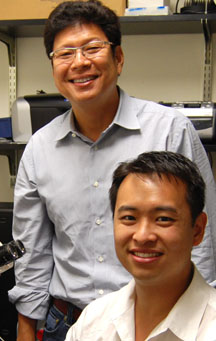
"This provides proof of concept for the medical treatment of this disease," says Professor Jerold Chun (back), who is shown here with first author of the new Science Translational Medicine paper Research Associate Yun Yung. (Photo by Cindy Brauer.)

Professor Jerold Chun speaks on this study.
A podcast from Science is available at http://stm.sciencemag.org/
content/3/99/99pc10.abstract
