

Team Led by Scripps Research Scientists Finds New Way that Cells Fix Damage to DNA
By Renee Twombly
A team of researchers at The Scripps Research Institute and other institutions has discovered a new way by which DNA repairs itself, a process that is critical to the protection of the genome, and integral to prevention of cancer development.
Scientists who study the repair of the DNA bases, which make up the information in the human genome, had known of only one type of method that cells use to fix a specific kind of damage to their DNA, but in the June 11, 2009 issue of Nature, the team found a novel way—one that combines elements from the known mechanisms and an unrelated second method that was previously not known to play a role in this type of DNA repair.
"We found a connection between the known DNA repair processes that people did not know was there," says Professor John Tainer, a member of the Skaggs Institute for Chemical Biology at Scripps Research, who led the study with Geoffrey P. Margison of the University of Manchester (United Kingdom) and Anthony E. Pegg of the Pennsylvania State University College of Medicine. "This changes the game, and gives us something important to look for in cancers that are resistant to chemotherapy."
This new mechanism is controlled by alkyltransferase-like proteins (ATLs), whose structure and function had been unknown and which had been identified only in bacteria and yeast. In addition to describing the function of ATLs, in the new study the scientists showed that ATLs exist in a multicellular organism, the sea anemone, which suggests this protein or its cousins in terms of repair activity also exist in other species, including humans.
Known Strategies for DNA Repair
Damage occurs to a cell's DNA on a continuing basis from outside sources, such as radiation and UV light, and from activities that go on day by day inside the cell. Most of this damage consists of damage to the DNA bases adenine, cytosine, guanine, and thymine. These bases pair up together inside the DNA double helix—adenine and thymine join together, and guanine and cytosine link to each other and their sequence forms the information in the human genome.
These bases can be chemically modified in a number of ways, including by alkylation, in which an alkyl group (or "adduct") is transferred onto a guanine base. When this happens, one of the hydrogen bonds holding guanine and cytosine together is removed, increasing the chances that thymine will be inserted across from guanine during DNA replication. If DNA is replicated with this "transition" error, a mutated gene results, so the information is changed. This can lead to harmful results, like cell death or cancer.
As shown in the reported work, this kind of damage occurs, for example, when chemicals derived from cigarette smoke stick to guanine, or when chemotherapy agents put an alkyl adduct onto guanine.
But that is where DNA repair mechanisms come in, which is good in the case of chemicals from cigarettes, but not so desirable when they repair genetic damage purposely induced by chemotherapy drugs intended to kill cancer cells.
The DNA repair process that removes such toxic "lesions" is known as base repair, and uses a protein called AGT (O6-alkylguanine DNA-alkytransferase) to remove the alkyl group before DNA replicates. The protein essentially sticks a chemical finger inside the DNA to flip the damaged guanine out from the DNA helix structure so that its adduct is exposed and can be transferred from the guanine to a part of its protein structure. The guanine is now repaired and can rejoin cytosine with three hydrogen bonds linking them.
AGT is believed to act alone, but there is another, unrelated repair process—nucleotide excision repair (NER)—that uses lots of proteins in its pathway. This repair occurs when bulky adducts stuck to bases distort the sleek shape of the DNA helix. Then a whole group of proteins come in and remove a patch of bases that includes the adduct, and DNA polymerase follows and fills in the patch while adding the correct base back.
A New Way
Before the new study, ATLs were believed to be involved in DNA damage responses, because they protected cells from DNA alkylation damage in lab experiments, but no one understood how they worked or what they did. In the new study, the team describes ATLs' role.
The scientists undertook a series of structural, genetic, and biochemistry experiments on the protein and determined its structure, both alone and with a guanine that had a methyl adduct and another with a smoking-derived adduct stuck on it. They found that the ATL structure looks like AGT. It, too, had a chemical finger that can rotate a damaged guanine base out from the DNA helix, but it doesn't remove the adduct like AGT does. Instead, ATL binds tightly to the damaged guanine and bends the DNA in a way that is more pronounced than what AGT does for repair.
"Base flipping by ATL is like a switch that activates the NER pathway, which then removes the alkyl adduct from the guanine," says first author Julie Tubbs, a research associate at Scripps Research. "So we believe that ATL is conceptually acting like a bridge, connecting the two DNA repair pathways—base and NER—together. This is a surprisingly general mechanism to channel specific base damage into the general NER pathway."
Before the new study, scientists also didn't know if ATLs functioned outside of single celled organisms. In the new study, however, the scientists discovered ATLs in two types of ancient organisms, archaeal bacteria and in sea anemone, suggesting this new bridging pathway may be general to most cells and organisms.
"What's especially important about these newly discovered ATLs is that we now know that ATLs exist in all domains of life, so it is very likely that ATL was common to the evolutionary branches before complex eukaryotes [single-celled or multicellular organisms whose cells contain a distinct membrane-bound nucleus]," Tainer says. "This suggests higher eukaryotes, including mammals and humans, will either have an ATL or have lost or replaced it with a protein of analogous function."
If ATLs are found in humans, Tainer sees that either inhibiting or bolstering their function could aid cancer therapy. Inhibiting DNA repair would help chemotherapy effectively destroy cancer cells. Augmenting ATL function could help protect sensitive tissue, such as bone marrow, that is easily destroyed during cancer treatment.
"There are all kinds of exciting ideas to emerge from this research," says Tainer. "For one thing, we now know what to look for when we see resistance to some chemotherapies."
In addition to Tainer, Margison, Pegg, and Tubbs, authors of the new study, "Flipping of alkylated DNA damage bridges base and nucleotide excision repair," are Vitaly Latypov, Amna Butt, Andrew Marriott, Amanda J. Watson, Barbara Verbeek, Gail McGown, and Mary Thorncroft of the University of Manchester; Sreenivas Kanugula of the Pennsylvania State University College of Medicine; Manana Melikishvili and Michael G. Fried of the University of Kentucky; Rolf Kraehenbuehl and Oliver Fleck of Bangor University; Mauro F. Santibanez-Koref of the University of Newcastle-upon-Tyne; Christopher Millington and David M. Williams of the University of Sheffield; Lisa A. Peterson of the University of Minnesota; and Andrew S. Arvai and Matthew D. Kroeger of Scripps Research. Tainer also holds a position at Lawrence Berkeley National Laboratory. For more information, see
http://www.nature.com/nature/journal/v459/n7248/abs/nature08076.html.
The study was supported by the National Institutes of Health, The Skaggs Institute for Chemical Biology, U.S. Department of Energy, the North West Cancer Research Fund, Cancer Research-UK, and CHEMORES.
Send comments to: mikaono[at]scripps.edu
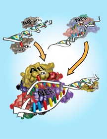
Members of the Tainer lab and their colleagues identified a previously unknown method that cells use to fix damage to their DNA—one that combines elements from two known mechanisms. Illustration by Mary O'Reilly. Click here to enlarge
