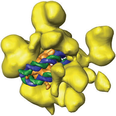
Macromolecular cryo-electron microscopy was used to calculate a 3D structure of the yeast RSC chromatin remodeling complex (in yellow) from images of thousands of individual RSC particles. Analysis of images recorded after incubation of RSC with nucleosomes revealed binding of the nucleosome (shown as a model with the histone octamer in orange and the DNA in green and blue) in a cavity of the RSC complex. Although RSC must use energy from ATP hydrolysis to remodel the nucleosome, binding alone seems to alter the nucleosome structure in a way that might faciliate ATP-dependent remodeling.