

Scientists Uncover New Mechanism Closely Linked to Neurodegeneration and Alzheimer's
Scientists from The Scripps Research Institute have uncovered a novel mechanism that may play a significant role in the development of Alzheimer's disease and a host of other neurodegenerative conditions. The discovery of this mechanism points towards potential new targets that could lead to treatments to enhance neuron survival.
The study was published in the November 11, 2008 edition (Volume 15, Issue 5) of the journal Developmental Cell.
The new study sheds light on the formation of large rod-shaped inclusion bodies that contribute to neurodegenerative injury and dysfunction. These rod-shaped assemblies, which are made up of the important protein actin (necessary for cell movement and division) and its key regulatory component cofilin, appear in abundance in animal models of neurodegeneration. These inclusions are especially abundant near amyloid deposits and neurofibrillary tangles in Alzheimer's disease.
"Actin/cofilin rods are abundant in brains of neurodegenerative disease patients, particularly Alzheimer's patients, and have long been suspected of playing a role in the progression of the underlying disease," said Scripps Research Professor Gary Bokoch, who conducted the study with Scripps Research postdoctoral fellow Timothy Huang and colleagues. "However, until our study, no one really knew how these rods were formed. Our study uncovered a mechanism for the formation of these rods during periods of energy stress that points to chronophin as an important new target for interrupting the process of neurodegeneration. It's a critical component of this pathway."
The brain, which represents just two percent of the body mass, uses 20 percent of the body's energy, making neurons highly susceptible to fluctuations in energy production from vascular or ischemic disturbances. The new study shows that chronophin, a cofilin-activating phosphatase, normally interacts with heat shock protein-90 (HSP90), a common molecular chaperone, to limit cofilin activation and consequent formation of cofilin/actin rods. When stress and/or cell damage depletes neurons of the nucleotide ATP, an important source of energy for the neuron, then chronophin is released from Hsp90 to become active, thus triggering the cofilin/actin rod-forming mechanism. The Hsp90-chronophin complex thus acts as an endogenous "biosensor" to detect energy depletion and invoke this (initially) protective response.
"Most researchers believe that when neurons undergo energy stress they try to preserve ATP—the energy currency of the cell," Bokoch said. "The polymerization and de-polymerization of actin, which is necessary for cell movement among other things, takes a great deal of energy, and that energy depletion initiates rod formation to help conserve ATP. Indeed, inhibiting actin turnover during periods of neuronal stress can reduce ATP depletion by as much as 50 percent, thereby preserving neuronal function."
Once formed, however, the actin/cofilin rods can remain in the cell for long periods of time, causing serious damage. For example, studies have shown that rod formation in neurons of Aplysia (sea slugs)reduces synaptic strength and neurotransmission. More ominously, studies in mice have shown these rods block transport of vesicles containing amyloid precursor protein, as well as enzymes involved in processing amyloid b, causing an accumulation of these precursor proteins associated with Alzeheimer's in the affected neurons.
The new study, however, reveals a key part of the process of forming the actin/coflin rods that might be targeted in future therapies.
"During ATP depletion, cofilin characteristically exhibits rapid dephosphorylation—removing a phosphate group—prior to co-assembly with actin into rods," said Bokoch. "Our study shows that chronophin is a critical prerequisite to this dephosphorylation process and, consequently, to increased rod formation."
The study shows that inhibiting or limiting the ATP-dependent HSP90 function appeared to simulate the effects of ATP-depletion by triggering cofilin dephosphorylation and rod assembly. These effects were dependent on chronophin, as the reduction of chronophin levels (by RNAi) largely prevented these effects. It is noteworthy that other studies have shown Hsp90 inhibitors to confer a level of neuroprotection in disease models.
Chronophin is also expressed in other tissues that are sensitive to ischemic injury such as the kidneys, and tissues that exhibit elevated ATP turnover, like the heart. This suggests that the chronophin/HSP90 mechanism may be involved in actin reorganization during ATP stress in these tissues. In cell types unable to form cofilin rods, chronophin may mediate cytoskeletal changes during energy stress, the study said, establishing this energy-sensitive mechanism as a way to directly link ATP stress with both homeostatic and pathological changes in the actin cytoskeleton.
In addition to Bokoch and Huang, other authors of Chronophin Mediates an ATP-Sensing Mechanism for Cofilin Dephosphorylation and Neuronal Cofilin-Actin Rod Formation, are Laurie S. Minamide and James R. Bamburg of Colorado State University. For more information, see http://www.cell.com/developmental-cell/abstract/S1534-5807(08)00399-7.
The study was supported by the National Institutes of Health, the American Heart Association, and Colorado State University.
For videos of actin/coflin rod formation from the Bokoch lab, see http://www.scripps.edu/news/press/images/20081110Bokoch/MovieS1.AVI and http://www.scripps.edu/news/press/images/20081110Bokoch/MovieS1.AVI.
Send comments to: mikaono[at]scripps.edu
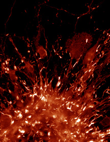
A new study from the Bokoch lab reveals a mechanism for the formation of actin/cofilin rods (shown forming in brain tissue, above), which have long been suspected of playing a role in the progression of Alzheimer's disease.
