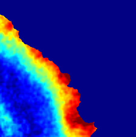
F-actin flow speed map computed from quantitative fluorescent speckle microscopy (qFSM) analysis of an epithelial PtK1 cell expressing active cofilin (GFP-cofilin S3A). Flow rates are color coded, ranging from fast flow in red to slow flow in blue. Increased cofilin activity induces a widening of the fast moving lamellipodium network at the cell leading edge. The study shows that Pak1-regulated cofilin functions as a spatial organizer of the lamellipodia and the lamella actin networks, whose interaction is required for efficient cell edge protrusion.