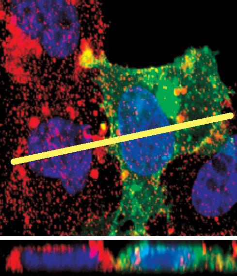
This image shows a confocal microscopy photograph of cells, in which blue represents cell nuclei, red represents MHC class I molecules, and green is a fluorescent protein indentifying cells that are expressing a protein from coxsackievirus B3. The yellow line in the upper panel shows the location where a "section" was cut through two cells, and the resulting side view is shown in the lower panel. In the lower panel's left-hand cell, MHC molecules can be seen distributed around the cell membrane, but in the lower panel's right-hand cell, which contains a viral protein, little MHC reaches the cell surface. (Image courtesy of the Whitton lab.)