
Quantum Leap
By Eric Sauter
A quantum dot (also called an artificial atom or a semiconductor nanocrystal) is, as the name implies, small—roughly the size of a human protein. In fact, if you lined up 3 million quantum dots of about seven nanometers each, they would only span the width of your thumb. Despite their small size, however, quantum dots cost more than $2,000 per gram and have captured the imagination of at least one popular author—Michael Crichton, who used a Hollywood version of such nanoparticles as biological terrorbots that threaten to take over the world in the novel Prey.
Phil Dawson, an associate professor in The Scripps Research Institute's Department of Cell Biology, isn't interested in world domination just yet, but he does want to find how quantum dots can be put to use imaging the activity of human proteases. Two new papers, one published in Bioconjugate Chemistry, the other in Nature Materials, offer an intriguing peek into this science that at first glance, perhaps even a second, belongs in the category of "strange but true."
Dawson, who graduated from Scripps Research's Ph.D. program, now the Kellogg School of Science and Technology, in 1996, did his postdoctoral work at California Institute of Technology and then returned to Scripps Research for good in 1997 ("I've been here forever," he said). He became interested in quantum dots a few years ago when he heard a lecture by Hedi Mattoussi, a researcher with the U.S. Naval Research Labs (NRL).
At that time, the big challenge with quantum dots was finding a way to combine the metallic properties of the dots with biological compounds so they could be used for imaging. With the publication of these two papers, that problem seems to have been addressed, and perhaps even moved ahead a dot or two.
Shining Example
Dawson and his colleagues have been intrigued by this topic because these luminescent quantum dots made from cadmium selenide and zinc sulphide (CaSe-ZnS) fall into a relatively new class of fluorescent probes that have, as the Bioconjugate Chemistry study concludes, "unique optical properties [that] suggest that they are superior to conventional organic dyes" for a range of biological applications.
The quantum dots were first coated with a biocompatible and water-soluble molecule (dihydrolipic acid), then mixed with a synthetic cell-penetrating peptide based on the HIV-1 TAT peptide that the virus uses to enter human cells. A tag on the peptide binds to the dihydrolipoic acid coating, and the whole thing self-assembles into a handy little nanosized complex ready to go work. The Bioconjugate Chemistry study allowed the researchers to examine the ability of these newly designed quantum dots to enter cells and to visualize their subsequent cellular localization.
"In our laboratory, we've always been interested in biophysical probes, and part of our work has been to incorporate unnatural chemical groups and place them inside proteins," Dawson said. "The NRL researchers wanted to hook quantum dots up to peptides and proteins. Consequently, we started working together. While these two papers used existing technologies to attach a number of different peptides to the surface of quantum dots, our goal is to eventually develop new ways to conjugate quantum dots to peptides."
The proteases the scientists want to test for (proteases are enzymes that breakdown proteins into peptides and amino acids) have a therapeutic and diagnostic bent—caspase-1, collagenase and thrombin. The first two are involved in cancer; and thrombin, with blood clotting.
"In the Nature Materials study, we attached a peptide to a quantum dot that could be cleaved by a specific protease to show activity," Dawson said. "On one end of the peptide we placed an organic molecule that has the effect of extinguishing quantum dot fluorescence. By attaching multiple peptides to each quantum dot, the majority of the fluorescence could be quenched. However, when the specific protease cleaves the peptide, the inhibiting group is no longer attached to the quantum dots. That's when the quantum dots shine on brightly."
Using quantum dots of different colors, each color linked to a peptide specific for one of the targeted proteases, the researchers created an assay that could image protease activity. They also demonstrated definitively that these particular quantum dots could be used in drug discovery to identify protease inhibitors. Focusing on thrombin, the researchers tested the inhibitory power of eight different compounds on thrombin activity. As it turned out, they got three potential "hits" that included several well-known thrombin inhibitors, including warfarin and sodium fluoride.
Next Steps
Even with this success, there are still some mysteries left on the table. The cell-penetrating peptides used to escort the quantum dots into the cell have been around for about the last 10 years, Dawson said, and are distinctive because of their relative lack of fussiness—they will take anything they are joined to into the cell. While there are a number of competing theories as to how they accomplish this rather astonishing task, nobody really understands how they work. In collaboration with Jim Delehanty at NRL, the Dawson lab showed the quantum dots localize largely to endosomes and also to the cytoplasm, but not into the cell nucleus.
Then there's the question of cytotoxicity. This issue has been studied by a number of researchers, including some at the University of California, San Diego, who reported in 2004 that the toxicity of the zinc sulphide-capped CdSe dots was "negligible," although the dots could be toxic in vivo under certain conditions. For his part, Dawson found that, in terms of acute toxicity, the results "were very promising," indicating that cells could be exposed to the these particular quantum dots with little or no problem.
The next step in the quantum dot scenario, of course, is using quantum dots in vivo, a move that remains somewhat problematic, at least in the short run.
"We would like to utilize the imaging power of quantum dots in vivo but we need to improve their solubility in biological systems," Dawson said. "Right now, because of their primary coating, the dots are highly acidic. As soon as pH levels veer towards neutral they tend to clump."
Which, as Dawson would be the first to point out, is not a good thing. So they are working to improve the bioavailability of quantum dots by modifying the coating and enhancing binding power.
"The dots have thousands of coating molecules on the surface," he said. "So far, we've been able to link over 20 cell-penetrating peptides on the surface of the quantum dots using something that could be compared to molecular Velcro. Now we're developing a monovalent but tighter link to bring them together."
Although it's something for the future, Dawson said, theoretically you could control the placement of more than a single type of peptide on the quantum dot surface. These flexibly faceted imaging tools would combine the advantages of cell-penetrating peptides with functionalized quantum dots for protease assays or targeting sequences highly useful in clinical diagnosis.
Would someone call Dr. Crichton's agent?
In addition to the members of the Dawson lab, authors of the new studies include members of the groups of Hedi Mattoussi, Igor Medintz, and James Delehanty at the Naval Research Labs, Washington, D.C.
The study was supported by the Office of Naval Research and the National Institutes of Health.
Send comments to: mikaono[at]scripps.edu

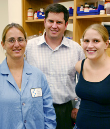
“In our laboratory, we’ve always been interested in biophysical probes, and part of our work has been to incorporate unnatural chemical groups and place them inside proteins,” says Associate Professor Phil Dawson (middle), pictured here with Research Associate Florence Brunel (left) and graduate student Theresa Tiefenbrunn. Photo by Kevin Fung.
