

In the Eye of the Be-Folder
By Jason Socrates Bardi
RNA folding has always been something of a celebrity's clumsy child—it's younger, much less glamorous, and perhaps destined to be overshadowed by its more famous relative: protein folding.
Protein folding is the queen of folding, and for decades, scientists have appreciated its beauty. Furthermore, scientists have been fascinated for decades by the mystery of protein folding—the question of how exactly the amino acid sequence causes proteins to adopt a single, stable three-dimensional structure that allows them to perform their specific biological functions in the body. This question is so important that it's been afforded a significant title: the protein-folding problem—beautiful and mysterious at the same time.
RNA folding, on the other hand, gets much less exposure. A recent Google™ search for the phrase "protein folding" turned up more than half a million separate web pages dealing in whole or in part with the subject, whereas a search for "RNA folding" yielded a mere 35,000 references.
Part of the reason for its relatively low profile is RNA folding's relative youth. It was only a generation ago that scientists even began to appreciate that RNA could fold and catalyze reactions. They had previously assumed that proteins were the only biological molecules that could carry out enzymatic reactions.
But in the last few years RNA folding has been coming into its own, and some of the most interesting questions in biophysics are emerging out of the field. Now, in a big breakthrough, a recent paper featured on a the cover of the journal Molecular Cell by Associate Professor Martha Fedor and her colleagues at The Scripps Research Institute answers one of the big questions in RNA folding: does the cellular environment contribute to how RNA molecules fold?
RNA Catalysis and Folding
Perhaps RNA folding was underappreciated for so long in part because there was little appreciation for what RNA could do once it was folded. Proteins, after all, have long been known to be the primary biological molecules that perform the catalytic functions underlying all basic functions in living cells and tissues. For years, nobody knew RNA could catalyze reactions as well.
That's because RNA is made up of nucleic acids, which are not nearly as versatile chemically as the amino acids that make up a protein chain. Furthermore, the idea that RNA catalyzes reactions didn't fit in with something known as the central dogma of molecular biology—that genetic information in the form of a single gene was transcribed into RNA and then translated into proteins.
The logic of the central dogma was that proteins were the end product of gene expression—the actors that express the words of the play. RNA, more of a stagehand than an actor, was simply the molecule that held up a genetic sequence so that it could be translated into a protein.
In 1982, however, a scientist named Tom Cech at the University of Colorado Boulder showed that RNA was the Rosencrantz and Guildenstern of the cell—minor characters, perhaps, but with an important role and interesting in their own right.
Cech made the completely unexpected discovery that RNA from a unicellular protozoan called Tetrahymena thermophila could perform auto-splicing. Cech shared the Nobel Prize in Chemistry in 1989 with Sidney Altman for his discovery of RNA molecules with catalytic function, and this opened up a whole new field for scientists: the structure, function, and mechanism of action of RNA enzymes, which soon began to be called "ribozymes."
In the last few decades, ribozymes have shown just how interesting RNA folding can be, and scientists have found a number of other cases where RNA folding leads to RNA-catalyzed activity in the cell. For instance, RNA refolding is known to play a role in the regulation of gene expression in bacteria. RNA secondary structure rearrangements are involved in the packaging of viral genomes into virions, or infectious particles. RNA structures are responsible for self-splicing in various fungi and bacteria. And some RNA are sensitive to metabolites in organisms and are involved in the metabolic control of gene expression.
However, a number of questions have remained about how RNA folding occurs in the body, some of which are only beginning to be addressed by scientists like Fedor, who studies RNA folding and catalysis as a member of the Department of Molecular Biology and The Skaggs Institute for Chemical Biology at Scripps Research.
Secondary Structures
The problem with studying RNA folding is that the quality that makes it interesting also makes it problematic—the ability to adopt multiple structures.
This is markedly different than protein folding. Proteins fold into their stable three-dimensional structures based on their primary sequences of amino acids. When a scientist takes a protein, and "denatures" or unfolds it, and then lets it refold, it will refold into the same structure as before. The reason for this, presumably, is that the folded structure is the most thermodynamically stable one.
RNA folding, on the other hand, presents a more complicated picture. RNA are able to form a large number of folded structures that are all more or less equally stable thermodynamically.
When biophysicists talk about protein structure, they are usually talking about both a protein's secondary structure (the way that a stretch of amino acids might form a helix, for instance), and its tertiary structure (the way a number of helices or other secondary structural elements collapse together into an overall three-dimensional shape). Protein secondary structure is not generally stable without the context of the greater three-dimensional structure.
But with RNA, it's not the overall tertiary structure that is stable but rather the secondary structure formed by the pairing of nucleic acids. RNA, after all, is a biological polymer made up of nucleotides. And, as in DNA, the nucleotides in RNA are able to pair through their complementary bases. With DNA, this pairing occurs between two separate but equal strands that line up. RNA, however, is typically single-stranded, so it pairs by looping back on itself, creating extremely stable secondary structures and allowing pieces of RNA such as ribozymes to freeze into place an arrangement of atoms. Some of these arrangements are chemically reactive, thus endowing the RNA with catalytic ability.
However, a large number of these alternative structures have no function. The dilemma for RNA, Fedor explains, is that many of these folded structures, including many structures that are non-functional, have similar stability. RNA can get stuck in what are known as kinetic traps, at least in model folding reactions in simple solutions. "They get hung up in these secondary structures," says Fedor, "because the [misfolded RNAs] are stable, they cannot unfold and refold."
And yet in living organisms RNA seems to be able to adopt a single, functional structure anyway—despite the similar thermodynamics of alternative structures. This then begs the question: how does RNA fold?
RNA Folding In Vivo
There are basically two possible answers to this question. Because RNAs are synthesized as linear polymers, RNA might fold up as they are being transcribed from DNA—something that has come to be known as the "sequential folding model," a favorite of some biophysicists for a number of years. The other possibility is that RNA folds only after it's completely transcribed, which can be referred to as "delayed folding model."
Wanting to examine the folding of RNA directly to compare the two models, Fedor and her colleagues analyzed RNA assembly during transcription in vitro and in vivo by monitoring the enzymatic activity of an RNA enzyme known as the hairpin ribozyme—a shortened, "minimal" form of catalytic RNA that's about 50 nucleotides long. This minimal hairpin ribozyme was discovered by botanists in plants infected by tobacco ring spot virus and a number of other viruses. It is part of what is known as plant satellite RNA.
Satellite RNAs are parasitic pieces of RNA that are not exactly viruses because they don't encode for proteins. Instead, they catalyze simple cut-and-paste reactions in order to replicate themselves, exacerbating or ameliorating diseases caused by plant viruses. A plant that has tobacco ring spot virus, for instance, will be more diseased if it also has this satellite RNA.
Since the hairpin ribozyme is so short, it's easily handled in the laboratory, which allowed Fedor and her colleagues to manipulate individual parts of the ribozyme and to study how it folds. The ribozyme has its own secondary structure, and certain bases are complementary to other bases downstream, which means that if the RNA happens to fold in half, these bases can stabilize the RNA into the hairpin shape.
The hairpin ribozyme also has the ability to self-cleave, which provides a good test for detecting that the folding occurs: if the scientists see the formation of the ribozyme, then folding was successful. Furthermore, by inserting upstream and downstream inhibitory structures (sequences of RNA that compete for the same complementary binding sites as the RNA that forms the hairpin), Fedor and her colleagues had a way of inhibiting the folding.
If the sequence folds correctly and forms the right complementary sequences, then the ribozyme forms and it cleaves itself in two. If, on the other hand, the RNA folds into an alternative structure, it will form a secondary structure without the ribozyme's catalytic activity and remain intact.
Significantly, this allowed the scientists to look at whether the folding occurred sequentially or after-the-fact by asking whether upstream inhibitory structures were more potent for inhibiting ribozyme assembly than downstream inhibitory structures.
The scientists expected that if the folding was sequential, the non-functional complementary RNA would appear first and would likely cause the RNA to fold into the nonfunctional form. A non-functional complementary sequence inserted at the tail end of the RNA would not be inhibiting because the ribozyme would already have a chance to form before the non-functional complementary sequence appears. The hairpin would form in the shortest possible time and self-cleave.
This is, in fact, is exactly what the Scripps Research team found in vitro. Putting the alternative complementary sequence close to the 5' end of the RNA wiped out most of the activity of the ribozyme, whereas inserting the alternative complementary sequences downstream at the 3' end had little effect.
And this is exactly what Fedor and her colleagues expected to find because these sorts of in vitro studies have been done in the past and have yielded the same results—which is one of the biggest pieces of evidence in favor of the sequential folding model.
However, Fedor and her colleagues had also developed a way to quantitatively measure the self-cleavage kinetics of ribozymes in yeast, and they wanted to use this technique to look for evidence of sequential folding in vivo. When they did, they found something that was completely at odds with the sequential folding model: the differential effect of inserting the alternative complementary sequence at the beginning or the end was lost.
"Downstream inhibitory structures were just as inhibitory as upstream inhibitory structures," says Fedor.
Not Consistent with Sequential Model
How are these conflicting data to be reconciled?
Fedor believes the best way is to abandon the sequential model altogether as something that occurs in the test tube but not in nature—in other words, to subscribe to the delayed folding model. These results provide enough evidence, says Fedor, to conclude that the sequential folding model cannot account for RNA folding in vivo.
But this raises another question: what features of the intracellular environment are delaying the folding of the RNA?
One possibility, says Fedor, are molecules known as chaperones—proteins that interact non-specifically with the RNA molecule's secondary structure and accelerate the interconversion of alternative structures or prevent them from folding until an entire region of RNA has been synthesized. Inside cells, misfolded RNA structures may be destabilized by RNA chaperones, which equilibrate the possible structures. So, no one fold has an advantage over another because it is expressed first. The chaperones are delaying the folding while the RNA is being expressed
These chaperones have been shown to work in vitro, but they have never been found in vivo. But the data obtained by Fedor and her colleagues, published in the July 1 issue of the journal Molecular Cell, show some evidence that chaperones or some other intracellular factors are at play during RNA folding.
Send comments to: jasonb@scripps.edu
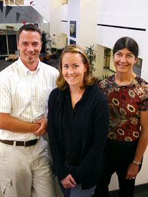
Research Associate Jason Harger, Research assistant Elisabeth Mahen, and Associate Professor Martha Fedor (left to right) have been investigating RNA folding.
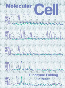
The recent paper was featured on the cover of Molecular Cell. Click to enlarge
