

Touching Molecules With Your Bare Hands:
Scripps Research Scientists Describe New Way of Interacting with the Unseen World of Proteins and DNA
By Jason Socrates Bardi
A group of scientists at The Scripps Research Institute has developed a new way of looking at and interacting with molecules so small that they cannot be seen even with the world’s most powerful microscopes.
The new technology, which combines hand-held objects with sophisticated computer displays, is called Tangible Interfaces for Structural Molecular Biology, and its creators envision it as a technology useful for both educational and scientific research.
“We want to be able to understand, communicate, and interact with complex structures in natural ways,” explains Molecular Biology Professor Art Olson, who led the research described in this month’s issue of the journal Structure. “The easier it is to hold a biological molecule in your hands, the easier it will be to figure out what it is doing in the body.”
By using cutting-edge three-dimensional fabricating printers that “print” solid objects out of thousands of layers of plaster or plastic, the group can construct models of proteins, DNA, and other tiny biological molecules. These models can be touched, twisted, tweaked, and tossed from person to person.
Then, using a simple digital video camera to capture and track images of these objects, the group is able to create an artificial environment in which the computer interfaces with the object in what is known as augmented reality.
The molecular model appears on the computer screen, tumbling and turning in real time as the person holding the object manipulates it, and software designed by the Scripps Research team enables the computer to superimpose scientific information about the molecule onto the display.
A Past Out of the Computer
Making physical models of objects like DNA and proteins may appear to be a step backwards in a sense because for years scientists have relied on computers to model complicated molecular structures.
To recall a time when computers were not the king of molecular displays, one has to step back more than a generation, before the advent of high-powered graphical processors cheap enough to go into any computer. In 1975, when Olson was finishing graduate school at the University of California at Berkeley, there simply was no way to interact with the structure of a molecule other than by making a physical model by hand.
This was an arduous task. It sometimes took months of painstaking work to glue the thousands of pieces of balsa wood or plastic together into a model. And when they were finished, many of these models were so fragile they had to include a support structure or be encased in a glass display so that they did not fall apart.
Olson remembers how he built his first protein structure by hand in a "Richards box"—which resembled something you would expect to find in a professional magician's kit rather than in a professional scientist's laboratory. A contraption in which a physical model of a molecular structure was built on a scaffold of metal rods with a half-silvered mirror that reflected hand-traced images of electron density, a Richards box revealed the molecular structure as a stick model (brass) of all of the protein atoms by fitting them into these electron density maps.
But times began changing in the 1980s as computer processors increased in speed and decreased in price. By mid-decade, machines powerful enough to display molecules on the screen had become inexpensive enough for laboratories to purchase.
These computers had two major impacts on molecular biology. One was that they were a great time-saver. The other was that the computers increased scientists’ ability to draw information from the structures of proteins and other biological molecules.
In fact, Olson was one of the leaders in the move towards computational molecular biology. He and his colleagues wrote a popular computer program called AutoDock in the 1980s, still an industry standard for predicting the interaction of ligands (drug candidates) with protein structures and other targets.
In the 1990s, computers had replaced physical models entirely, and a graduate student working on structural biology would probably never have laid eyes (much less hands) on a physical model—sitting instead for hours in front of a computer monitor. And much of the software that powered these computer models was not simple to operate. Whole graduate courses were devoted to learning a single piece of software, and many students had to become experts in programming as well as biology.
A few years ago, Olson began to renew his interest in some of the old ways. Rather than rely solely on computers and the flat digital molecules they display, he wanted to go back to constructing and manipulating physical models. He knew that such models would help scientists understand the molecules even better.
Luckily, technology has once again caught up, and a whole generation of new, “solid” printers have greatly increased the ease with which molecular models can be constructed. Instead of months of painstaking work to construct these models, they can now be printed automatically, and put together in a matter of minutes.
Olson and his colleagues have designed innovative ways to construct the pieces of a structure, such as a protein. They have been experimenting in making hybrid models, for instance inserting magnets to snap together two pieces of a model. This has allowed them to demonstrate such processes as viral assembly, the three-dimensional folding of a long amino acid chain into a compact protein, and docking between two proteins.
On the Interface of Technology and Biology
Olson showed a recent visitor to his laboratory a couple of models that demonstrated the utility of the new printers. His calling card is a walnut-sized model of a spherical virus that opens up to reveal a plaster bon bon that looks deceptively sugary and has his laboratory’s name and web site imprinted across the spherical surface.
And that is just the beginning.
To demonstrate self-assembly—a common process whereby tiny objects like viruses put themselves together from small identical subunits into a compact structure, sort of a three-dimensional puzzle—Olson puts pieces of plastic that look like highly complicated Legos™ about the size of a quarter into a jar and shakes them up.
The pieces represent protein molecules that come together to form a virus particle, and tiny magnets imbedded in them help orient them in the correct way. After some vigorous shaking, there is an assembled model of a virion inside the jar.
It’s a neat trick, but his next trick is even neater: using these “tangible” models as a computer interface. These models are not meant to be mere knickknacks or objets d’art. They are intended to be actual research tools.
By turning back to the computer and implementing a software tool called augmented reality, he is able to mix his real-world objects with computer-generated graphics where the computer interfaces with the object.
The manipulation is not just a parlor trick, he explains, but is linked to computation. He demonstrates by taking a model of a protein and displaying on the computer screen its electrostatics—blue and red clouds surrounding the object he is holding that show favorable and unfavorable interactions. As he brings two ends of the molecule in close proximity to each other, the cloud surrounding the ends changes from glowing blue to glowing red. The protein doesn’t “like” to have these two ends so close to one another.
Similarly, says Olson, such tangible interfaces could be used to manipulate models and predict molecular interactions.
Moving back to his models, he shows that not everything he works on is completely finished. Showing his visitor another, larger plastic bottle filled with multi-colored plaster chips that are supposed to fit together, he shakes it wildly.
“With such a large model,” he explains, “it takes a little bit longer.” Shake, shake. Some of the fragments come together. Shake, shake. Others are still trying to find their way into the growing ball. Shake, shake. After a few minutes, he stops. There are still some pieces that are unassembled in the bottom of the jar.
“This is one we are still trying to design,” says Olson.
To read the article, “Tangible Interfaces for Structural Molecular Biology” by Alexandre Gillet, Michel Sanner, Daniel Stoffler, and Arthur Olson, see the March, 2005 issue of the journal Structure (13, 483–491) or go to: http://dx.doi.org/10.1016/j.str.2005.01.009
This work was supported by the National Institutes of Health and the National Science Foundation.
Send comments to: jasonb@scripps.edu
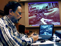
"The easier it is to hold a biological molecule in your hands, the easier it will be to figure out what it is doing in the body," says Professor Art Olson. Photo by Kevin Fung.
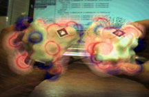
Manipulating 3D printed molecular models, scientists explore protein interactions via augmented reality. Image courtesy of Cell Press.
