

Stem Cell Therapy Holds Promise for People with Retinitis Pigmentosa
By Jason Socrates Bardi
A team of researchers from The Scripps Research Institute was able to preserve visual function in mice that were genetically predisposed to developing a profound degenerative disease that destroys their retinas.
The team injected adult bone marrow-derived stem cells from mice or humans into the back of mouse eyes at an appropriate stage of development and these injections dramatically curtailed retinal degeneration. In the latest issue of the Journal of Clinical Investigation, the team shows that the treated eyes of the mice, when compared to the fellow untreated eyes, had a completely normal retinal vasculature, had significantly improved retinal tissue, and responded to light.
"The surprising findings by Dr. Friedlander and colleagues establish a dramatically new paradigm for understanding and potentially treating retinal degenerative diseases using a cell-based approach," says Paul A. Sieving, director of the National Eye Institute, National Institutes of Health. "Determining the precise mechanism of this cell-mediated rescue presents an exciting research challenge and is a high priority for the Institute."
This approach could potentially be used to treat disorders of the retina that have vascular and neuronal degeneration. Such inherited degenerative retinal disorders are known collectively as retinitis pigmentosa. According to the National Eye Institute, more than 100,000 Americans suffer from retinitis pigmentosa, which is caused by more than 100 different genetic mutations.
"For patients with retinitis pigmentosa, this may be tremendously important," says Martin Friedlanderwho led the study. "In the mouse, we have used both mouse and human cells to preserve nearly normal vascular and neuronal architecture of the retina in a disease model that ordinarily exhibits profound degeneration. Our hope is that if these results translate into humans with the disease, we would be able to maintain vision for these patients longer."
Currently, there is no way to treat patients with retinitis pigmentosa and no way even to slow the disease, says Friedlander, who is an associate professor in the Department of Cell Biology and the chief of the Retina Service in the Division of Ophthalmology, Department of Surgery, at Scripps Clinic. Friedlander has had a longstanding research program looking for new and better ways of treating eye diseases such as age-related macular degeneration, diabetic retinopathy, and retinitis pigmentosa. Four years ago several members of his group, including Atsushi Otani and Michael Dorrell, began to explore the potential utility of adult bone marrow-derived stem cells for the treatment of these disorders, and a series of studies published over the past three years has confirmed that such an approach may be useful both scientifically and clinically.
The Key to Treating Retinitis Pigmentosa
The retina is akin to an extension of the brain into the back of the eye. It is a layer of blood vessels and nervous tissue that covers about two-thirds of the back of the eyeball and is connected to the brain through the optic nerve. Its primary purpose is to capture light and transduce those physical cues into electrical signals, which it then sends to the back of the brain where the sensory signals are interpreted into the visual experience.
Retinas contain a number of specialized cells, including the rods and cones, which capture light and send electrical signals to the brain, and glial support cells. The retina also has an extensive vasculature—a fine mesh of blood vessels formed in the third trimester of human gestation and in the first month postnatally in mice by endothelial cells, the major cell type lining blood vessels.
In normal mice, these retinal blood vessels form during the first three to four weeks after birth and provide blood to the inner two thirds of the retina. In mice that are predisposed to developing retinal eye disease, the outer layer of the retina containing the rods and cones as well as other neuronal layers degenerate within a few weeks after birth. Most of the cell death occurs by apoptosis, or programmed cell death. The retinal blood vessels are present in three layers, and in the model of retinal degeneration the two deeper retinal vessel layers completely degenerate by about one month after birth.
In humans with retinitis pigmentosa, a very similar process occurs, and this leads to profound vision loss and eventual blindness. The disease starts as progressive night blindness and the gradual loss of peripheral vision, and it leads eventually to complete tunnel vision or in some cases total blindness.
More than 100 different types of gene mutations have been documented that lead to this degeneration of the retina, and about one in every 3,500 people suffer from loss of vision caused by retinitis pigmentosa.
But at least in mice, Friendlander and his colleagues found that blindness can be prevented by injecting adult bone marrow-derived stem cells into the back of the eye.
A New Kind of Protection
The group's basic approach starts with selecting what are called lineage negative stem cells from the bone marrow. Adult bone marrow stem cells are "pluripotent" and have the potential to develop into a number of different cell types, such as red blood cells, platelets, or white blood cells. Lineage negative stem cells have the capability, among other things, of becoming endothelial cells—the major type of cell that lines the body's blood vessels.
Friedlander and his colleagues found that the lineage negative stem cells, once injected into the mouse eye, would localize to a type of star-shaped glial support cells called astrocytes. During prenatal human development, astrocytes guide endothelial cells into place where they can proliferate and form blood vessels.
Later in life, under certain circumstances, astrocytes will proliferate and can do the same thing, acting as beacons to bring the stem cells to the retinal vasculature. These stem cells were guided by the retinal astrocytes to the vasculature in the back of the eye. There some were incorporated into the vasculature while others took up positions very close to the blood vessels; both were able to survive—in fact, they seemed to be protected from death.
Once at sites of the retinal vasculature, the stem cells would provide a protective effect, rescuing and stabilizing the retinal vessels when they would otherwise degenerate.
Significantly, the injected stem cells protected the retinal neurons from death at the same time. The neuronal protection seemed to be specific for the cones, the types of photoreceptors found predominantly in the human macula, the center of the retina responsible for fine, or reading, vision.
Friedlander and his colleagues investigated the molecular basis of this process. It turns out that these stem cells are loaded with a type of protein known as "heat shock proteins."
These cells are sort of like firefighters, says Friedlander. They are protected from apoptosis the way that a firefighter is protected from heat and flames by specialized gear. And the stem cells extend their protection to the surrounding cells of the retina, much as a firefighter, by protecting one position, might protect an entire area of a burning building.
Once inside the retina, these stem cells produce their heat shock proteins and probably induce other cells to produce them as well, thus preventing the retinal and vascular degeneration ordinarily observed in this mouse models of retinitis pigmentosa.
The next step, says Friedlander, would be to perform additional preclinical studies aimed at determining dosage and possible toxicities of a treatment based on this research and then taking the approach into clinical trials.
"The clinical paradigm is novel and, frankly, we were very surprised at the results," says Friedlander. "Our data in two mouse models of retinal degeneration suggest that it may be possible to use autologous bone marrow-derived stem cell grafts to provide a dramatic vasculo- and neurotrophic protective effect in a variety of retinal degenerative diseases including RP and macular degeneration."
However, warns Friendlander, as encouraging as it is that they were able to achieve the same rescue effect in mice with human bone marrow cells, the technique is still some distance from the clinics. At the moment, Friedlander and his colleagues are continuing their efforts with human bone marrow-derived stem cells.
"These cells are truly remarkable and provide a rationale basis for using vascular reconstructive approaches in the treatment of diseases in which the endogenous vasculature is subject to degeneration or malfunction," he says. "Since most diseases that cause profound visual loss have abnormalities in the vasculature, the potential clinical application of this approach is quite broad."
The research article "Rescue of retinal degeneration by intravitreally injected adult bone marrow-derived lineage negative hematopoietic stem cells" was authored by Atsushi Otani, Michael Ian Dorrell, Karen Kinder, Stacey K. Moreno, Steven Nusinowitz, Eyal Banin, John Heckenlively, and Martin Friedlander and appears in the September 15, 2004 issue of the Journal of Clinical Investigation. The JCI articles can be accessed at http://www.jci.org.
The research was supported by the National Eye Institute, the Robert Mealey Program for the Study of Macular Degenerations and the Kovner Family Fund.
Send comments to: jasonb@scripps.edu
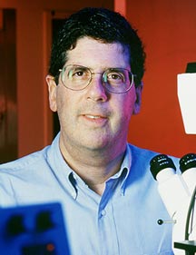
Associate Professor Martin Friedlander hopes that his latest research will lead to treatments to help maintain the vision of patients with retinitis pigmentosa.
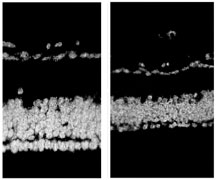
Stem cells from human bone marrow show promise for reducing retinal degeneration. Figures show an eye predisposed to degeneration treated with stem cells (left) and the contra-lateral eye in the same model treated with control cells.
