| |
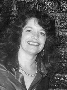
Heart Cells' SuicidesBy Jason Socrates Bardi The influences of Roberta Gottlieb's early days growing up on a cattle ranch in rural New Mexico continue to be meaningful in her life today as an associate professor in the Department of Molecular and Experimental Medicine (MEM) at The Scripps Research Institute. One influence of those days on the open range is the sense of self-reliance and creativity they instilled in her. She recalls how her father once saw a metal and wood device for immobilizing calves in order to brand them at a cattle show and came home and built the same sort of thing from scratch. And the endless expanse of the high desert was fertile ground for thinking creatively. "You ended up coming up with a lot of things on your own," she says, adding that this is something that she finds absolutely necessary in the cutting-edge world of basic biology. The other way that the rancher's life prepared her for work in the MEM's Division of Hematology is that she used to get up every morning before sunrise in order to tend to the herd. Today, she has no herd, but rising early is still de rigeur to attend to her laboratory. Apoptosis in the HeartAt Scripps Research, Gottlieb and her colleagues study an area of emerging importance in the biology of heart disease—the process of apoptosis in heart cells. Apoptosis, also called programmed cell death or cell suicide, is a methodical process whereby cells take deliberate steps to achieve their own demise. Apoptotic cells express proteins that break down their DNA, DNA repair enzymes, and structural proteins like lamins and actin. The morphology of a cell changes as it undergoes apoptosis, shrinking away. Despite how dramatic this may sound, apoptosis is a calm sort of death in contrast to necrosis, another major form of cell death in the heart that is characterized by sometimes violent cell lysis and inflammation. When Gottlieb came to Scripps Research in the early 1990s to work in the laboratory of MEM Professor Bernard Babior, she was interested in looking at apoptosis in the immune cells known as neutrophils. She had finished her medical degree several years before, and had completed her residency and fellowship in pediatric oncology, and a postdoctoral fellowship in molecular biology at the University of California, San Diego. At that time, scientists assumed that the death of heart cells after ischemia was by necrosis. While a visiting scientist in the Babior lab, cardiologist Robert Engler wondered whether apoptosis might also be taking place. Gottlieb joined the team and applied a newly-described method of detecting apoptotic cells in situ, and they soon were the first team to demonstrate that apoptosis occurred in the heart after ischemia/reperfusion. Apoptosis, it turns out, is a big issue in heart attacks, which are the number one killer in the United States. According to the National Heart, Lung, and Blood Institute, about 12.6 million Americans suffer from coronary heart disease, the most common form of heart disease. This disease often leads to an acute myocardial infarction, the technical term for a heart attack. Some 1.1 million Americans suffer heart attacks each year, and approximately 515,000 of these are attacks are fatal. Currently, the main treatments for heart attacks address the initial thrombus or blockage to the artery in order to restore blood flow to the heart. Doctors use thrombolytic "clot busting" drugs to dissolve the blockage chemically, or angioplasty—tiny balloon catheters often followed by a wire mesh stent—to mechanically prevent the artery from collapsing. Over the long term, myocardial infarction leads to fibrosis, the formation of scar tissue that replaces dead heart tissue. Heart attack survivors often have weakened hearts because this scar tissue cannot function properly. These patients often require additional procedures, such as the insertion of pacemakers or heart transplants. The blockage starves the heart tissue of oxygen, triggering a whole cascade of events. However, tissue damage may continue to worsen in the hours following the attack, even after the clot is gone. In fact, the restoration of blood flow triggers additional injury due in part to dramatic shifts in intracellular ion concentrations, formation of damaging oxygen radicals, and inflammation. Additional damage occurs because the ischemia may have made the heart cells trigger their own deaths through apoptosis. "As you restore blood flow," says Gottlieb, "you create a number of changes that are deleterious and can trigger cell death." Reperfusion is necessary for patients who have heart attacks, for if you do not do it, the heart cells will die from necrosis and the patients will not live. The irony of restoring blood flow in a blocked artery after a heart attack is that the reperfusion may cause some additional damage, although it is still preferable to leaving the heart muscle without blood flow permanently. Acid DrainIn a heart that has been cut off of its oxygen supply from an ischemic blockage, the heart cells do what they can to survive. Cells that do not have enough oxygen begin to perform glycolysis, the breakdown of glucose, in order to supply energy in the form of a molecule called ATP. But glycolysis generates lactic acid as a by-product, and so ischemia leads to a drop in the intracellular pH (the cells become more acidic). Facing the accumulation of lactic acid, the cells try to normalize their pH by pumping the acidic molecules out of the cell. This works, but it also can trigger the exchange of ions in and out of the cell. Normally, cells maintain distinct "gradients" of ions like sodium, potassium, and calcium, and these differences in concentration of ions on the inside and outside of cells establish chemical and electrical driving forces that can be used to perform a range of basic cellular operations. But when ions like sodium and calcium are exchanged following ischemia, they might be exchanged contrary to their normal concentrations. Sodium ions, for instance, are normally kept low inside cells, but following reperfusion, the intracellular concentration of sodium goes up. So does calcium. In order to deal with the increase in calcium, heart cells rely on their mitochondria. Mitochondria are the highly complex organelles located inside virtually every cell in the human body that normally supply cells with most of the energy they need. But during ischemia, the mitochondria try to reduce the levels of calcium inside the cell by sequestering it. This influx of calcium into the mitochondria triggers a response that results in the catastrophic release of proteins from within the mitochondrial membrane. "This makes it difficult for the cell to recover because [without functioning mitochondria], the cell doesn't have an energy synthesis machine," Says Gottlieb. Worse, the ruptured mitochondria also release factors that trigger apoptosis, through activation of proteins that break down proteins (called caspases) and others called endonucleases, which chew up DNA in the cell. Another event that happens in the mitochondria following reperfusion is the production of what are referred to as "reactive oxygen species." These are compounds like hydrogen peroxide and oxygen radicals that are highly reactive and can react with lipids and proteins in the mitochondrial membrane to produce oxidized lipid and protein molecules. These can also compromise a cell's function. "It becomes a very complicated set of events that are going on simultaneously—like a three-ring circus," says Gottlieb. "Any one of [these events] alone can result in the death of the cell." Because of the potential for damaging the heart with reperfusion, many doctors and scientists have been looking for ways to intervene at the beginning of reperfusion. One of the approaches that scientists like Gottlieb have taken has been to look for so-called "drugable" targets. These are proteins or other biological molecules that are involved in some part of a biological phenomenon like a heart attack that might be amenable to therapy. Finding drugs that effectively do this is another matter, but the first step is to find the targets. One such target, says Gottlieb, is a common protein called cytochrome p450. This is actually a family of several dozen metabolic enzymes that are involved in activities like removing toxins from the body. Cytochrome p450 proteins seem to be important for heart attacks as well. Increases in the expression of cytochrome p450 proteins have been correlated with some of the more traditional risks factors for heart attacks—like high cholesterol, smoking, and diabetes. And cytochrome p450s have the ability to take a fatty acid that is generated during ischemia and convert the acid to signaling molecules called eicosanoids. "These interact with a whole host of different enzymes inside the cell," says Gottlieb. While all the effects of the signaling molecules are not known, what is known is that they impact a number of different pathways involved in ion transport and mitochondrial function and start a cascade of events that are lethal to heart cells. The activity of cytochrome p450s could potentially be critical to whether a cell survives or not. Because of this, cytochrome p450 proteins seem to be a good drugable target, and inhibiting them might improve the survival of heart attack victims. Gottlieb and her colleagues have been evaluating drugs that are selective against particular cytochrome P450 proteins to see how they perform in models of ischemia and reperfusion. An Exciting New ResultIn a recent issue of the journal Proceedings of the National Academy of Sciences, Gottlieb describes how a common antibiotic called chloramphenicol, which inhibits protein synthesis in mitochondria, can also reduce the size of a myocardial infarction and also reduce the production of these reactive oxygen species. Gottlieb and her colleagues report that they could not detect an effect of chloramphenicol on protein synthesis in mitochondria, suggesting that its protective effect was not due to this. Instead, they propose that chloramphenicol protects the heart by inhibiting cytochrome P450 proteins. To support this, they demonstrated that two other cytochrome P450 inhibitors, which were known not to inhibit protein synthesis in mitochondria, also reduced the size of a myocardial infarction and the production of these reactive oxygen species. "This is a therapy that could possibly be applied after the ischemic insult (heart attack)," says Gottlieb. Other individuals in her laboratory are looking at questions related to what happens inside mitochondria during apoptosis, more specifically, how apoptosis may be relevant in the heart. Some lab members are studying a fatty lipid molecule that is produced by platelets and by a variety of tissue cells called sphingosine 1-phosphate, which binds to cellular receptors called the S1P receptors, activating them and regulating a range of physiological functions that include cardiovascular function and blood pressure. This work is in collaboration with Scripps Research Professor Hugh Rosen, and is an effort to determine if sphingosine 1-phosphate is an important pathway in the heart and whether compounds that are chemically similar to the lipid or drugs that bind to its receptor might play a role in heart attacks as well. Working with Scripps Research Institute President, Professor Richard A. Lerner, and with Scripps Research Associate Professor Paul Wentworth, Jr., Gottlieb and members of her laboratory are trying to determine if ozone is formed during myocardial ischemia and reperfusion, and whether this dangerous form of oxygen is responsible for the damage to lipids and proteins in the heart. In another avenue of research, Gottlieb and her colleagues are trying to apply a new technology called "protein transduction domains" for delivering proteins into cells as a way of studying signal transduction in the beating heart. A protein transduction domain is a short sequence of amino acids that enables an entire protein to enter the cytoplasm of a cell. She and her colleagues are using these sequences to drag anti-apoptotic proteins into cells to see if they can protect the heart. Into the SunsetIf Gottlieb's days are busy with experiments, grant and paper writing, and meetings, her evenings are even busier with her children entering the teen years—just old enough to have lots of places to be carted off to but not quite old enough to drive themselves. Gottlieb gets up early every day so that she can come home and get her children fed, shepherd them to their various after-school activities, and get them to bed so she can get a few hours of sleep herself in order to be up early the next day. This is the fun part, she admits, and she enjoys activities like taking Tae Kwon Do with her son. "He was having so much fun that I decided to join," she says. "Then I got hooked on it." Soon, she says, she will test for her black belt. Surely such hand and foot skills must also owe something to her days back on the ranch in New Mexico. Asked if she can still lasso a cow from atop a horse at daybreak, she laughs. "We never did that," she says, "but I know how to vaccinate one."
|
|
|