Nano-Origami:
Scientists Create Single, Clonable Strand of DNA That Folds into an Octahedron
By Jason Socrates Bardi
A group of scientists at The Scripps Research Institute has designed,
constructed, and imaged a single strand of DNA that spontaneously folds
into a highly rigid, nanoscale octahedron that is several million times
smaller than the length of a standard ruler and about the size of several
other common biological structures, such as a small virus or a cellular
ribosome.
Making the octahedron from a single strand was a breakthrough. Because
of this, the structure can be amplified with the standard tools of molecular
biology and can easily be cloned, replicated, amplified, evolved, and
adapted for various applications. This process also has the potential
to be scaled up so that large amounts of uniform DNA nanomaterials can
be produced. These octahedra are potential building blocks for future
projects, from new tools for basic biomedical science to the tiny computers
of tomorrow.
"Now we have biological control, and not just synthetic chemical control,
over the production of rigid, wireframe DNA objects," says Research Associate
William Shih of Scripps Research.
Shih led the research, described in the latest issue of the journal
Nature, with Professor Gerald Joyce of the Department of Molecular
Biology and The Skaggs Institute for Chemical Biology at Scripps Research.
Compartments and Scaffolds on the Nano-Scale
Similar to a piece of paper folded into an origami box, the strand of
DNA that Shih and Joyce designed folds into a compact octahedron—a
structure consisting of twelve edges, six vertices, and eight triangular
faces. The structure is about 22 nanometers in overall diameter.
These miniscule octahedral structures are the culmination of a design
process that started one day when Shih was building a number of shapes
with flexible ball and stick models in the laboratory. This exercise attracted
his attention to an important structural principle: frames built with
triangular faces are rigid, while cubes and other frames built with non-triangular
faces are easily deformed.
Translating this principle to a scale over a million times smaller,
Shih sought to design a DNA sequence that would fold into a triangle-faced,
and therefore very rigid, object. Shih and Joyce settled on trying to
build an octahedron. Shih and Joyce constructed a 1669-nucleotide strand
of DNA that they designed to have a number of self-complementary regions,
which would induce the strand to fold back on itself to form a sturdy
octahedron. Folding the DNA into the octahedral structures simply required
the heating and then cooling of solutions containing the DNA, magnesium
ions, and a few accessory molecules. And, indeed, the DNA spontaneously
folded into the target structure.
The researchers used cryoelectron microscopy, in collaboration with
Research Assistant Joel Quispe of the Scripps Research Automated Molecular
Imaging Group, to take two-dimensional snapshots of the octahedral structures.
Significantly, the structures were highly uniform in shape—uniform
enough, in fact, to allow the reconstruction of the three-dimensional
structure by computational averaging of the individual particle images.
Potential Applications
Shih and Joyce note that because all twelve edges of the octahedral
structures have unique sequences, they are versatile molecular building
blocks that could potentially be used to self-assemble complex higher-order
structures.
Possible applications include using these octahedra as artificial compartments
into which proteins or other molecules could be inserted—something
Joyce likens to a virus in reverse, since in nature, viruses are self-assembling
nanostructures that typically have proteins on the outside and DNA or
RNA on the inside.
"With this," says Joyce, "you could in principle have DNA on the outside
and proteins on the inside."
The DNA octahedra could possibly form scaffolds that host proteins for
the purposes of x-ray crystallography, which depends on growing well-ordered
crystals composed of arrays of molecules.
Another potential application is in the area of electronics and computing.
Computers, which rely on the movement and storage of charges, can potentially
be built with nano-scale transistors, but one of the big challenges to
accomplishing this is organizing these components into integrated circuits.
Structures like the ones that Shih and Joyce have developed might someday
guide the assembly of nanoscale circuits that extend computing performance
beyond the limits set by silicon integrated circuit technology.
The article, "A 1.7-kilobase single-stranded DNA that folds into a nanoscale
octahedron" was authored by William M. Shih, Joel D. Quispe, and Gerald
F. Joyce and appears in the February 12, 2004 issue of the journal Nature.
This work was supported by the National Aeronautics and Space Administration,
The Skaggs Institute for Research, the National Institutes of Health through
the National Center for Research Resources, and through a Damon Runyon
Cancer Research Foundation fellowship.
Send comments to: jasonb@scripps.edu
Go back to News & Views Index
|
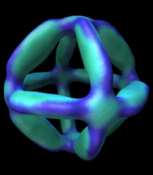
An image of a clonable DNA octahedron, roughly the
size of a small virus, visualized using cryo-electron microscopy and single-particle
reconstruction analysis. False colors indicate relative electron density.
Image courtesy of Mike Pique.
|