Persistent LCMV infection rendered immature Schwann cells defective in myelin formation
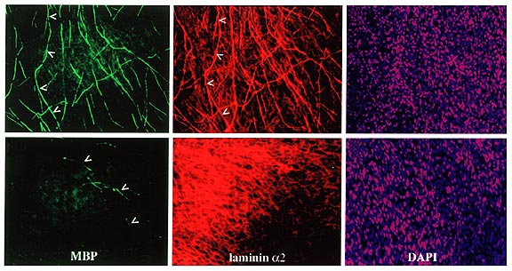
After 21 days, both infected and control cultures were labeled to detect the myelination (left column), laminin-2 organization (middle column), and nuclei (right column). In the left column, anti myelin basic protein antibodies were used to detect myelination (green), which is markedly reduced in the infected cell culture. In the middle column, anti-laminin antibodies were used to show the laminin (red), which appears disorganized in the infected cell culture. In the right column, labeling with Hoechst dye shows the nuclei (purple) of both the infected and control cells. Note how there is little difference between the two—signifying that that the nuclei of the infected cells are still intact.
Image courtesy of Proceedings of the National Academy of Sciences.