| |
Blood Flow Beneath a Microscope
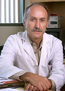
"I'm going to show you the best way to visualize platelets," says Professor Zaverio Ruggeri, giving an impromptu lesson in blood clotting last month in a darkened laboratory in the Molecular and Experimental Medicine (MEM) building at The Scripps Research Institute (TSRI). Ruggeri holds the Roon Chair in Cardiovascular Research and heads MEM's Division of Experimental Hemostasis and Thrombosis. He also directs TSRI's Roon Center for Research in Arteriosclerosis and Thrombosis, endowed in 1981 with private funds from the Roon family—Leo and Anna Miesem Roon, their son Donald Roon and daughter-in-law Lois Roon. The Roon Center supports researchers in thrombosis and arteriosclerosis, and sponsors an annual lecture by a prominent scientist in the field. Ruggeri continues his demonstration, showing how an all-day video can record the results. For the last several years, he has done similar experiments in this room, the Roon Research Laboratory in Artheriosclerosis and Thrombosis, three or four times a week. "I have thousands of hours of video," he smiles. The videos show the movement of blood cells over a flat surface under the microscope. In particular, they show the movement of platelets—those flat, molecule-filled cytoplasmic disks in the blood that are necessary for clotting. On the surface of the microscope stage is a chamber over flows which blood exposed to proteins, cells, and other materials. The surface is engineered to mimic the surface of a blood vessel—with a layer of endothelial cells on top of collagen and other "matrix" components. Ruggeri uses these systems to look at the interaction of platelets with various surfaces encountered in the circulation, and to study the interaction of individual platelets with the endothelial cells and wounds. Above the noise of the cooling fans, Ruggeri points out how the computer scans slices of the sample flowing over the surface in successive one-micron slices, from the matrix material on the bottom, through the layer of endothelial cells, and finally above the endothelial cells where the platelets are flowing and accumulating. To mimic a flesh wound, an artificial wound has been made on this surface by scraping away some of the endothelial cells, exposing the matrix components underneath. In real life, the exposed endothelial matrix affects the flow of the platelets, ultimately leading to clotting of blood. Here, Ruggeri is able to monitor and record these microscopic events. "We'll see how the platelets interact with this 'lesion'," says Ruggeri as he starts the flow, "where they are accumulating in relation to the cut." There, on the screen are cascading blips not much larger than a single pixel. Those blips, Ruggeri says, are single platelets. As they rush over the scarred surface of the artificial wound and encounter the subendothelial layer, they clump into bright spots several micrometers across (composed of thousands of platelets). The platelets are clotting. "These are very dynamic events, and we are interested in [capturing] the real-time dynamic aspects of them," says Ruggeri. The Two Faces of ClottingScientists are interested in the molecular mechanisms that govern clotting because many individuals suffer from diseases related to these mechanisms, such as bleeding disorders. The most famous such bleeding disorder is hemophilia. Ruggeri, however, has spent most of his career studying a disorder that is more common but less-well-known: von Willebrand disease—a genetic defect caused by mutations in a large, sticky protein called von Willebrand factor, which interacts with platelets to initiate clotting. Von Willebrand disease is less severe than hemophilia. It is, however, the most common hereditary bleeding disorder, affecting at least one percent of the population. Another reason scientists are interested in clotting is its potential for therapeutic intervention in vasculature disease. Clotting is an essential physiological process, but at the same time, the blood components that heroically stop bleeding, also nefariously cause diseases such as heart attacks and stroke, which are the most common causes of death in the United States today. Platelet adhesion and clotting are central to acute and chronic arterial diseases, since platelets have a predominant role in initiating acute adhesion in coronary arteries. For many years, people like Ruggeri have been trying to design a way to prevent the von Willebrand factor protein from initiating platelet adhesion and clotting in the hopes of ameliorating arterial disease. "As yet, there is no small molecule inhibitor," says Ruggeri. "But my prediction is that in the future, there will be a specific anti-platelet drug targeting this interaction." Ruggeri is contributing to this effort by elucidating the molecular mechanisms of these physiological processes. Recently, in collaboration with TSRI Staff Scientist Reha Celikel and TSRI Associate Professor Kottayil Varughese, he published a paper in the journal Science describing the structure of a platelet receptor called glycoprotein Ib bound to the coagulation protein thrombin, which induces blood clotting by producing the sticky protein fibrin. "We are trying to understand, at the structural level, how glycoprotein Ib, thrombin, and von Willebrand factor could assemble into complexes that influence one another," says Ruggeri. "There are some interesting points of convergence."
Ruggeri came to TSRI from Milan in 1978 for what he thought would be a brief, one-year stay as a research associate. At that time, he was a practicing physician who specialized in hematology, particularly in treating patients with hereditary bleeding disorders like von Willebrand disease. "I came here to acquire knowledge that I could apply to techniques to improve the diagnosis of these patients," says Ruggeri. He soon became interested in the mechanism of von Willebrand disease and wound up staying an extra year before returning to Milan. When he did return in 1980 and began working with patients again, he found it impossible to ignore the scientific implications of what he saw. As associate director of the Hemophilia and Thrombosis Center at Policlinico Hospital in Milan, Ruggeri noticed that the von Willebrand factor proteins behaved differently in different patients with different forms of the disease. Von Willebrand factor, the largest protein in human plasma, is a polymeric protein that is produced by endothelial cells that line blood vessels. One subunit of von Willebrand factor is more than 2,000 amino acids, and dimers of these subunits form the building blocks of the polymer. The polymers are large chains of various numbers of these dimers, and the size of these polymers is related to the function of the protein, which is to mediate the attachment of platelets to areas in the circulation where there is a lesion. The larger the polymers, the more sticky they become. To induce blood clotting, the polymers should be as long as possible, since the number of binding sites increases with length (something biologists refer to as multivalency, with each subunit of a multimer contributing an equal number of binding sites). Ruggeri observed that, in some patients, the von Willebrand factor was not as large (and sticky) as it needed to be. In others, it grew as large as it needed to induce blood clotting, but it was hyper-vulnerable to attack by a blood protease and was degraded too quickly. And sometimes, the von Willebrand factor proteins in patients seemed to have multiple problems. These differences in the proteins translated into differences in how the platelets behaved—for instance, how they stuck to one another, and how they stuck to the surfaces of blood vessels. "The tests we were doing in [the patients] were showing different results, and it was clear that there were different categories of the disease," he says. Point of No ReturnRuggeri returned to TSRI in 1982, armed with these observations and determined to examine the causes at the molecular level. In the years since, he has made many important discoveries related to the disease and its various phenotypes—which, it turns out, are caused by mutations in different parts of the von Willebrand factor molecule. Prior to the arrival of modern molecular biology and genomics, this sort of work relied largely on characterizing phenotypes and performing molecular studies that were, by today's standards, crude. Ruggeri and others purified the von Willebrand factor protein from plasma and digested it with enzymes into its various domains. With the advent of molecular biological techniques that enabled investigators to express domains of a protein in large amounts, by the early 1990s several laboratories, including Ruggeri's in collaboration with TSRI Associate Professor Jerry Ware, began a line of work that has yielded several high-resolution structures of von Willebrand factor domains. Ruggeri, in collaboration with Celikel and Varughese, solved the first such structure, the A1 domain of von Willebrand factor, in 1999. This was an arduous task as the A1 domain was difficult to crystallize. As a protein, von Willebrand factor is naturally sticky, which leads to trouble with solubility. But once Ruggeri and his colleagues crystallized and solved this first domain, similar studies with mutants and other structural variants became easier. The A1 von Willebrand factor domain has a core of beta strands surrounded by a number of alpha helices. It appears to be identical to domains used by a whole family of other proteins—intergrins, for instance. In von Willebrand factor, the A1 domain is crucial for blood clotting because it interacts with platelets. Not surprisingly, many of the mutations that have been reported in von Willebrand factor protein from patients with the disease are in this domain. Gain of Function—In a Bad WayRuggeri describes how one mutation to the A1 domain von Willebrand factor protein causes von Willebrand disease. Paradoxically, this mutation does not knock out the function of the domain, but causes it to become more active. The mutation causes the A1 domain to bind to platelets too tightly. Normally, von Willebrand factor is essential for bringing platelets to lesions on the surface of vessels through which blood is flowing rapidly. In order to do this, the proteins need to bind to both the platelets and to the collagen and other components of the matrix, which are exposed when a vessel is cut. "But," says Ruggeri, "they are only meant to interact [with platelets] where there is a lesion." The gain-of-function von Willebrand factor mutations cause the circulating von Willebrand factor proteins to bind to the platelets avidly, even when the platelets are in normal circulation. The platelets, in turn, become coated with the von Willebrand factor protein, and thus exhaust the sort of long, sticky, multimeric polymers needed to induce clotting. In the end, patients bleed because the platelets don't stick. "They fly over the surface," says Ruggeri. Ruggeri notes that several other mutations in von Willebrand factor function in different ways to cause the bleeding disorder. Such mutations can make the protein lose the ability to multimerize or to bind to the platelets at all. The FutureKnowing the molecular bases of the various causes of von Willebrand disease is extremely helpful in designing therapies. Patients who have the protein, but in whom it is not released in the blood can be treated with a peptide after they are cut. This peptide forces their cells to release stores of the von Willebrand factor protein and corrects the bleeding problem. This type of treatment is worthless for patients with different kinds of mutations. On the other hand, patients who have a malfunctioning form of the protein per se are best treated by the administration of normal von Willebrand factor isolated from donated blood. Asked about the future, Ruggeri notes that there is still a lot to do in the present. Today, he points out, even though the A1 domain and another domain of the von Willebrand factor protein have been solved, these account for only about one third of the total protein. No structure for the entire protein monomer exists and no structure for the dimer exists either. "There is still a lot of work to do," says Ruggeri. "We are slowly building up our knowledge."
|
|
|