Scientists Report Cryo-EM Structure of a Human Platelet Integrin Molecule
By Jason Socrates Bardi
Two researchers at The Scripps Research Institute (TSRI) recently published
the first detailed three-dimensional model for the human platelet integrin
alphaIIbbeta3—a signaling molecule that is
important for activating platelets, which leads to the healthy formation
of blood clots in response to a cut as well as clots that obstruct blood
flow to healthy tissue.
The structure, which was obtained through electron cryo-microscopy (cryo-EM),
image analysis, and molecular modeling, will appear in this week's issue
of the journal Proceedings of the National Academy of the Sciences
and reveals new structural details of this important molecule.
These results will be relevant for the design of new drugs to treat
health conditions in which the formation of blood clots is undesirable,
such as during myocardial infarctions (heart attacks) and strokes. Medical
techniques like balloon angioplasty and intracoronary stent implantation
are designed to clear blocked arteries but can also cause the formation
of thrombi. In fact, several recent clinical trials have demonstrated
that alphaIIbbeta3 inhibitors have benefit in the
medical treatment not only for heart attacks but also during angioplasty
and stent placement.
The Structure Revealed
Picture an integrin as a large button on an overcoat. Most of it sits
exposed on the outside of the coat, but it is connected by threads that
extend to the inside of the material.
Actually, the integrin is something of a scientific marvel because it
transduces signals over a distance of nearly 200 Ångstroms, whereas
most signaling molecules work over a distance of 10 to 15 Ångstroms.
Integrins are made up of two separate polypeptide chains (called the alpha
and beta chains) that come in a variety of forms. Recent crystallographic
studies by the Arnaout group at Harvard revealed that the large extracellular
portion of the molecule, the "button," has 12 distinct folding "domains."
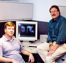
"Integrins have a large and very complicated structure," says Mark Yeager,
M.D., Ph.D., who published this latest integrin structure with his postdoctoral
fellow Brian Adair, Ph.D. "They are a broad class of signaling molecules
that affect diverse biological processes such as development, angiogenesis,
wound healing, neoplastic transformation, and thrombosis."
The integrin alphaIIbbeta3
is particularly important in the process that leads to thrombosis because
it is one of the key signaling molecules on platelets, the disk-shaped
cells that are involved in blood clotting. There are typically 40,000
to 80,000 on the surface of any given platelet, spanning the platelet
membranes, where they are involved in signal transduction—detecting
specific molecules (ligands) outside the cell and communicating that detection
to the inside, or vice-versa. The ligands are proteins attached to other
cells, in the extracellular matrix, or that freely circulate in the bloodstream.
When these ligand molecules bind to the extracellular integrin subunits,
they induce "outside-in" signaling in which a three-dimensional conformational
change at one end of the integrin is propagated through the membrane to
the other end of the integrin. Thus, the binding event on one side of
the cell is "transduced" through the cellular membrane. Proteins inside
cells can also bind to the cytoplasmic "threads" of the integrins and
alter the extracellular affinity for ligands, a process termed "inside-out"
signaling.
According to the model that Adair and Yeager now propose, the alphaIIbbeta3
integrins have multiple conformations and undergo dramatic shape changes
depending on whether the molecule is in the high- or low-affinity state.
The "threads" that transmit the signal through the membrane are folded
as a coiled-coil of alpha-helices.
When the platelet is activated, the integrin is in a "high-affinity"
form, extending far out on the outside of the cell and exposing its binding
site to potential ligands. One of the ligands that binds to the high-affinity
conformation is fibrinogen, a circulating blood protein that can bind
integrins at both ends. Fibrinogen is present in large amounts in the
blood, and when platelets are active, the high-affinity integrins bind
to fibrinogen proteins, which in turn bind more integrins at their other
end, and this leads to the formation of massive clots of platelets.
"We hypothesize that the switch between the high- and low-affinity states
for the integrin involves flexing at hinge-like connections between certain
domains in the extracellular subunits so that the molecule collapses into
a tighter overall structure," says Yeager. "It is a very dramatic event."
A Suspect Jackknife
Though scientists have known for many years that integrins are important
in many physiological processes, detailed structural information on these
molecules has been elusive.
The size of the integrins, and the fact that they span the membrane
confounded structural studies of the proteins. In fact, the only way to
solve the structure was to chop off the membrane-spanning regions and
solve the individual parts separately by x-ray crystallography.
But until recently, there were no high-resolution structures even of
these extracellular domains. Then the Arnaout group published the crystal
structure of the domains in the journal Science about a year ago.
However, this structure showed that the ligand-binding head region was
bent back, like a jackknife, to the point where it was almost touching
the region of the protein that would connect the transmembrane "threads".
"The crystal structure provided a lot of new insight," says Adair, "But
it does not seem that this 'jackknife' form is the major conformation
for the intact molecule."
How the Technique of Electron Microscopy Works
The first electron microscope was built by Ernst Ruska in 1933, for
which he received the Nobel Prize in 1986 at age 80. Electron microscopes
use magnetic lenses to bend a beam of electrons to image tiny objects,
similar to the bending of light by glass lenses in a light microscope.
EM looks at a range of magnifications, from no more than an ordinary light
microscope that magnifies up to 60 times to those that magnify up to 1,000,000
times.
TSRI is one of the few centers in the world with an integrated program
in electron microscopy of biological complexes and macromolecular machines.
The Center for Integrated Molecular Biosciences is directed by Ron Milligan.
Two other Scripps scientists, Bridget Carragher and Clint Potter, were
recently awarded an NIH Research Resource Grant to develop automated molecular
microscopy. Adair and Yeager used the Philips/FEI microscopes at CimBIO
to collect their data.
Cryo-EM, which is the technique used in the current study, requires
that samples be spread in a thin film and then frozen on a copper meshwork
grid. The freezing process occurs in a few milliseconds at about a million
degrees a second. In this way the frozen water is in a glass-like vitreous
state, which is an excellent environment to preserve biological molecules
in near-physiological conditions—a significant advantage over x-ray
crystallography, where the proteins are often crystallized in pieces and
in exotic buffers.
Adair and Yeager purified the integrin molecules from human platelets
in mild detergent solutions that mimic the oily environment of the platelet
membrane.
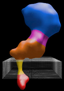
The computational challenge was to sort out thousands of different views
of the integrin molecules and combine them to derive a 3-D map. The map
revealed the overall shape and size of the entire integrin, including
the large extracellular domain, the small cytoplasmic domains and the
transmembrane coiled-coil.
Adair and Yeager then used the EM structure as a "molecular envelope"—like
a mold, into which the 12 domains derived by x-ray crystallography could
be docked. By this combined approach a detailed description of the structure
and action of complicated molecular machines such as integrins can be
derived.
The article, "Three-dimensional model of the human platelet integrin
alphaIIbbeta3 based on electron cryomicroscopy and
x-ray crystallography" is authored by Brian D. Adair and Mark Yeager and
appears in the October 29, 2002 edition of the journal Proceedings
of the National Academy of Sciences.
This work was supported by the National Institutes of Health, the National
Heart, Lung, and Blood Institute, and a postdoctoral fellowship from the
California affiliate of the American Heart Association (to Adair). During
the course of this work, Yeager was an Established Investigator of the
American Heart Association and is now the recipient of a Clinical Scientist
Award in Translational Research from the Burroughs Wellcome Fund.
Go back to News & Views Index
|