Delivery of ATP-Raf to Tumor-Associated Blood Vessels Causes Endothelial and Tumor Cell Apoptosis
(A-C) Athymic WEHI models were subcutaneously implanted with M21-L melanoma, and tumors were allowed to grow to ~400 mm3. Models were then given a single i.v. injection of avb3-NP-Raf(-). Controls were injected with the avb3-NP coupled to a shuttle vector. After 24 or 72 hours, the tumors were resected, fixed, sectioned, and stained.
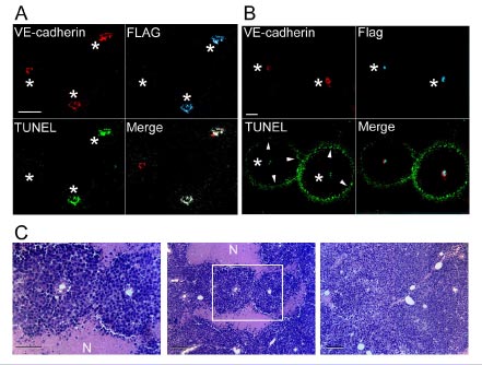
(A)
Tumors harvested 24 hours after treatment were immunostained
for VE-Cadherin (endothelial cells), FLAG (gene expression), and TUNEL
(apoptosis) (bar=50 microns). Asterisks denote blood vessels.
(B)
Tumors harvested 72 hours after treatment
were stained as above (bar = 50 microns). Arrowheads denote ring of tumor
cells undergoing apoptosis.
(C)
Tumors harvested 72 hours after treatment with avb3-NP-Raf(-)
(left and center panel) or controls (right panel) were stained with hematoxylin
and eosin. Necrotic tissues are denoted by N (bar = 50 microns, left panel
and 100 microns, center and right panel).
Reprinted with permission from Science. Copyright 2002 American Association for the Advancement of Science.