|
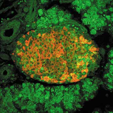
| The picture shows a section of a pancreas from an in vivo model harboring pancreatic beta cells lacking responses to interferons. The model was infected with coxsackievirus B4 (CVB4) and sacrificed on day 3 post infection. FITC (green) channel shows cells stained for CVB4 using an antibody directed against to VP-1, a capsid protein antigen in CVB4. Rhodamine (red) staining show insulin positive beta cells. Virus is present in the exocrine part of the pancreas. Because they lack responses to interferons, the beta cells in this model become infected with virus (seen as the presence of virus within beta cells in the islets of this model) and will within days develope Type 1 diabetes as a result of beta cell loss. Picture by Malin Flodström, Dept of Immunology, and Brian Smith, The Core Microscopy Facility, TSRI. |