| |
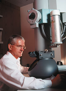
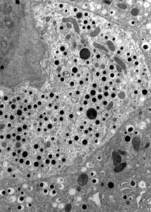
Focusing on the Electron Microscopy FacilityBy Mika Ono Malcolm Wood, director of TSRI's Electron Microscopy Facility, adjusts the focus, contrast, brightness, and magnification of the black-and-white image of a blood vessel on a screen, looking for clues to a problem of excess bleeding. Still not satisfied, he moves on to see if he can glean more insight from the next sample. "The ability to see and to understand what you are seeing is critical in microscopy," says Wood. "Experience does help interpreting the images, as does the confidence to interact with the equipment. You can't just accept the first image on the screen as real." Many scientists at TSRI send samples to the Electron Microscopy Facility to draw on the expertise of Wood and his staff, microscopists Theresa Fassel and Brian Smith. Assistant Professor Henrik Ditzel, for one, wanted to identify the location of several proteins on neutrophils, a kind of white blood cell highly destructive of microorganisms, to help understand a type of autoimmune reaction. "Malcolm was very helpful," says Ditzel. "It's great to have high-quality core facilities like this one on campus. They make the progress of research much faster." The Electron Microscopy Facility, which participates in 15 to 20 TSRI research projects a month, houses two electron microscopes in the basement of the Molecular Biology/Skaggs Institute Building and a confocal and a deconvolution microscope in the basement of the Stein Building. Projects are turned around in three days to two weeks or more, depending on the requirements of the job and the number of samples provided. Electron microscopy, a technique developed in the 1930s, can magnify a sample theoretically to the angstrom level, a level at which individual molecules can be detected. To prepare a sample for electron microscopy, cells are fixed and embedded in resin for support, then cut into extremely thin slices—70 nanometers thick—using a diamond knife. The sections are then picked up on a mesh grid and stained with heavy metals such as uranium or lead for contrast. In the electron microscope, the various lenses are electromagnets that essentially manipulate the beam of electrons in a manner comparable to the glass lenses of the light microscope. In the transmission electron microscopes, the beam of electrons passes through the sample and the electrons impact the phosphorescent screen to create the image of the sample. "Essentially, in electron microscopy you are looking at a shadow of the parts of the cell that have differentially taken up heavy metals," explains Wood. "In this shadow, you can identify structures." Although electron microscopy offers extremely high magnification and better resolution than the light microscope, preparing samples for this technique can be a time-consuming process. "In immuno-electron microscopy, it's often a balancing act to find a concentration of chemicals that will preserve cells without destroying antigenicity," Wood says. "In light microscopy, sample preparation is more straight-forward. Although the magnification it offers is not as high, light microscopy has an important role to play as a guide for further research." In addition to its drop-off service, the Electron Microscopy Facility offers an open-access program in which researchers can learn to use the microscopes and go on to work with them independently. Training, which is conducted by the facility's staff, usually lasts 10 to 12 hours for electron microscopes, 8 to 10 hours for light microscopes. "People are sometimes surprised when they use the instruments for the first time," says Wood. "In general, you don't see the images shown in a textbook. More often, the structures appear at an unexpected angle or plane. You have to think three-dimensionally while looking at two dimensions." For more information on the Electron Microscopy Facility, contact Wood, x4-8186 or e-mail mwood@scripps.edu.
|
| |