Rendering of ILE pocket
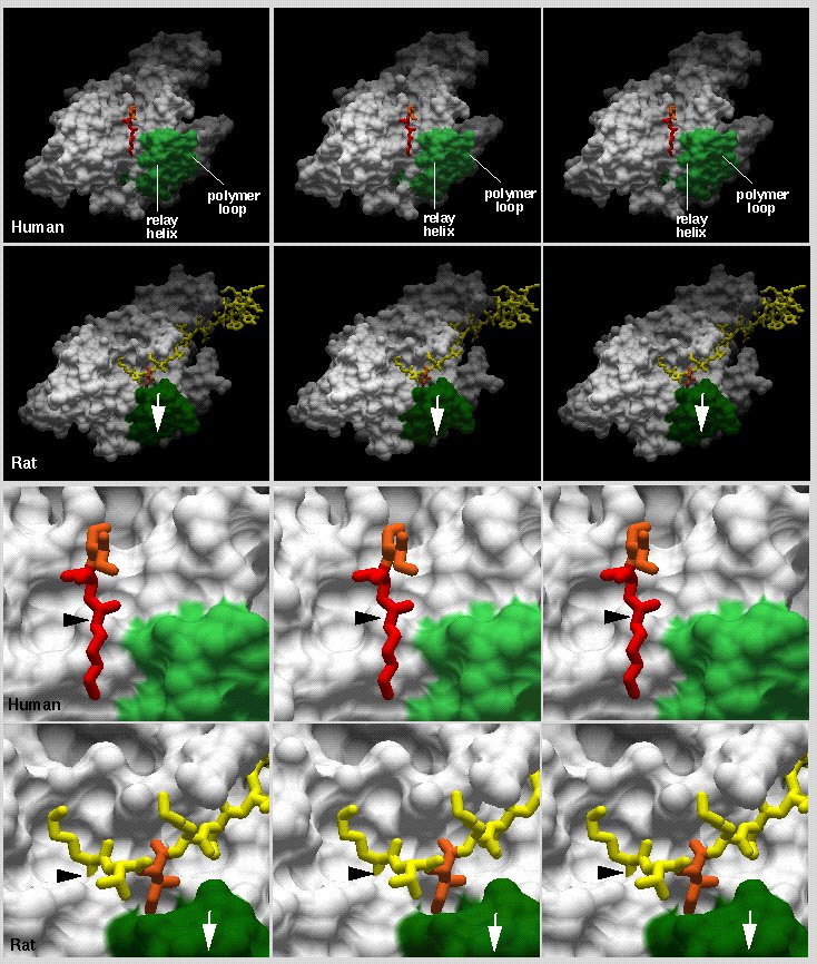
Shown here are cross-eyed (left pair of trio) and wall-eyed (right pair)
stereos of the human and rat kinesin structures. Based on arguments outlined
in the article, we propose that these two
structures represent the ADP/nucleotide-free and ATP/ADP-Pi states of kinesin
respectively. In the ADP/nucleotide-free state (human), the neck linker
is mobile and only a small portion of it is seen in the x-ray structure (red
and orange). ATP binding to the active site causes the relay helix and polymer
loop to move (white arrow) from the downstroke position (light green) to the
upstroke position (dark green), creating a pocket on the surface of the
motor core. Insertion of I325 (orange, I327 in rat kinesin)
into this pocket triggers zippering of the neck linker onto the core. In the
docked rat kinesin neck linker (yellow), pockets for additional residues can
also be identified, e.g. a conserved N329, two residues to the right of I327.
The black arrowhead indicates where the visible portion of the neck linker
originates on the rendered surface.
Acknowledgements:
Human kinesin PDB# 1BG2
Rat kinesin PDB# 2KIN
Brain Sheehan made the renderings, using AVS modules written by Mike Pique.
Abel W Lin composed the final illustration.
contents © 2000
awl