Open the 'TEM direct control' dialog with a click on . The 'Tem direct control' dialog
is a so-called non-modal dialog, i.e. it remains open as a parallel dialog.
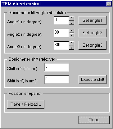
'TEM direct control' dialog
Goniometer tilt angle (absolute)
This section can be used for a quick access the tilt of the goniometer. You can enter 3 often used tilt angles either directly
by typing the value in the corresponding edit fields or with the spin control .
A click on one of the Set angle X sets the corresponding tilt angle.
Goniometer shift (relative)
This section can be used to move the specimem along the x and/or y axis in units of µm . Therefore enter the desired shift
distances into the appropriate edit fields and click on Execute shift.
Position snapshot
A click on opens the 'Take / Restore snapshot' dialog
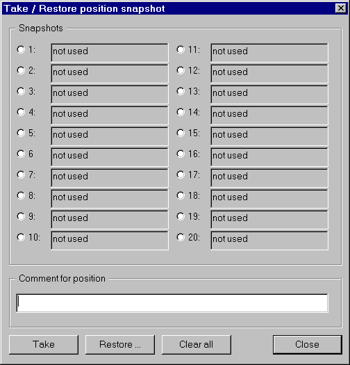
'Take / Restore snapshot' dialog
To "store" a position, enter a text into the edit field in section 'Comment for position', then select one of the 20 snapshot positions
and click . Now all goniometer data, magnification, spotsize, intensity,
beam shift X/Y and image shift X/Y are "snapshooted" and stored under the selected snapshot position.
To "restore" a position select one of the up to 20 used snapshot positions
and click . Now all goniometer data, magnification, spotsize, intensity,
beam shift X/Y and image shift X/Y are restored.
A click on clears all snapshot positions.
6.4.2 Control gonio ...
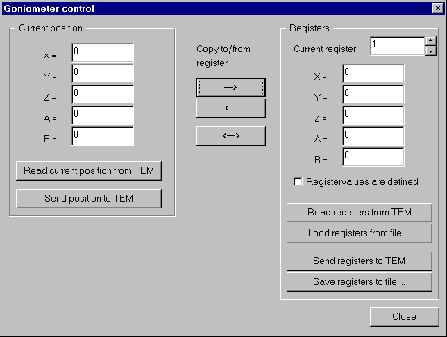
Pressing opens the 'Goniometer control' dialog.
The 'Goniometer control' dialog
The 'Current position' section shows the actual values of the goniometer after they are transferred from the microscope to the workstation. You can also enter specific values for the position and send them to the microscope.
The 'Registers' section shows the actual values for the chosen register (edit box 'Current register') after they are transferred from the microscope to the workstaion or loaded from a file.
The option Registervalues are defined defines whether a register parameter set ( X, Y, Z, A and B ) is really ( or should really be ) valid
within the EM.
The three buttons between the two sections of this page do the following actions:
copy current position to register
copy register to current position
swap current position and register
6.4.3 Autofocus ...
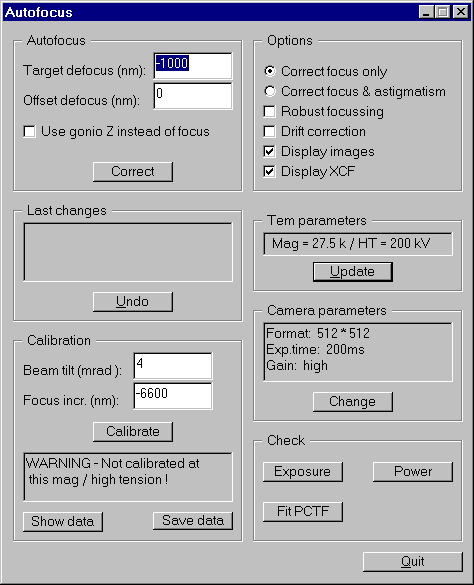
The 'Autofocus' dialog is started with .
The 'Autofocus' dialog
This page is divided into 7 sections.
Autofocus
In this section you can enter your desired defocus in nm. It is also possible to enter a value for an offset defocus.
An offset defocus is useful if the starting defocus is close to zero defocus.
The option 'Use gonio Z instead of focus' will not change the objective coil currents to change the focus,
but use the goniometer's z-axis to focus. All the temporary focus changes (e.g. offset defocus ) will still be performed using the objective lenses.
Options
The options 'Correct focus only' and 'Correct focus & astigmatism' are used either to calibrate/correct only the defocus or to calibrate/correct
both defocus and astigmatism. 'Robust focussing' uses a ten times smaller tilt to ensure that the image shift is not that much as with the normally
suggested tilt.
Note: a smaller shift results in a smaller accuracy for calibration/correction.
'Drift correction' takes more images to determine and correct the estimated drift that occurrs during calibration/correction.
'Display images' shows all the exposures taken during calibration/correction on the screen.
'Display XCF' shows all crosscorrelation functions that are computed during calibration/correction.
Last changes
If there have already been changes the last change is visible here. The last change can be undone.
TEM parameters
In this section the currently used TEM magnification is shown. Update here if the magnification is changed.
Calibration
In this section you can change the suggested beam tilt and focus increment for the calibration in the corresponding edit fields.
Note: choose the values such, that the tilted images still correlate. As soon as the tilted images do not correlate correctly,
the result of the calibration will become nonsense and is therefore useless for the correction.
If there is already a calibration available for this magnification, the date of the calibration as well as x-to-y tilt ratio and the angle are shown.
Good values for the ratio are near to 1.0 and for the angle are about ±90.0° (or ±270.0°).
Section 'Camera parameters':
In this section the camera parameters for calibrating/correcting focus and/or astigmatism are shown.
Note: due to memory reasons, the number of pixels read from the camera must not exceed a quarter of the maximal size.
E.g. if the maximal format is 1024x1024 pixels, the maximal autofocus format is 512x512 pixels. Binned or not binned makes no difference,
except for large focus changes where a large field of view is required to measure the beam tilt induced displacement.
Check
This section contains tools to check whether the correction of focus and/or astigmatism was performed successful or not.
Important: The calibration of autofocus and autoastigmatism correction need the TEM being properly aligned., e.g. the beam tilt pivot points and the rotation center must be aligned.
Here is a recipe how to calibrate.
Set the display format to 512 viewport size
When starting the calibration routine you will be asked if you are close to focus. This is a crucial point. If the initial focus setting is too far away from focus, the measured displacement will be too large or the image disappears when a beam tilt is applied. To avoid these problems we suggest to proceed as follows with an initial calibration. (A recalibration is always necessary if a new column alignment was made.)
Use a high contrast specimen for the calibration; e.g. amorphous carbon film with graphite or gold particles.
Choose a medium magnification to start with (30k)
Focus the specimen to zero defocus as good as you can
Mark the following options: 'Correct focus only', 'Display images' and 'Display XCF'
Start the calibration now by pressing the button 'Calibrate'.
If you have already focused the image press 'YES' when the question 'Close to focus' appears on the screen, otherwise lower the screen and focus again. Several images are now acquired and displayed together with the crosscorrelation function of images with opposite beam tilt. To control the process, check if every image displayed shows a normal image (even with some slight astigmatism) and if the correlation peaks are within 2/3 of the correlation plane.
After successful calibration use the autofocus function now to adjust focus precisely to zero defocus. To do so you have to set the Target defocus to '0' and the offset defocus to a decent value (e.g. for magnification of 30k => offset defocus should be around -3000 nm; this value varies for different magnifications). Now press the correct button. after successful focusing you can see the focus change in the field 'last changes'.
Now focus should be very close to zero defocus. Check with the 'exposure' and 'Power' button.
Now you can redo the calibration either with the option 'correct focus only' or the option 'correct focus & astigmatism' on.
After successful termination save the calibration parameters for this magnification with the 'Save Data' button.
Now you can go up or down by 1 step in magnification (click the 'update' button or use the 'exposure' button to update the magnification setting in the computer). Continue with the calibration for all other magnifications. If you have focussed to zero defocus at the previous magnification, you can do the calibration now in one step.
General remarks:
If a focus measurement starts at a defocus close to zero, the displacement of the images with opposite beam tilt is very small. This will result in a less precise measurement. To overcome this problem, you can define an 'offset-defocus'. This will produce a larger displacement and thus a more precise measurement.
For Philips CM series TEMs, resetting the defocus in the EM-Page (on the microscope) after focusing to zero, allows control of the relative focus changes.
For further information on 'Beam_tilt-Induced-Displacement Autofocus' see also A.J.Koster and W.J. de Ruijter, Practical autoalignment of transmission electron microscopes, Ultramicroscopy 40 (1992) 89-107.
6.4.4 Drift ...
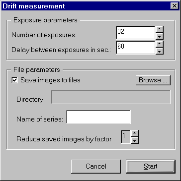
Use to open the 'Drift measurement' poupmenu:
The 'Drift measurement' dialog
In the section 'Exposure parameters' are two input fields for the number of exposures and the delay time between two exposures.
The section 'File parameters' is active, if the checkbox 'Save images to files' is marked. If it is, then you must select a file to store the results before the measurement is started.
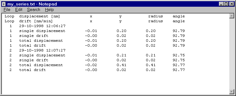
If you have decided to save the image file, then the results of the measurement are also stored to a file (in the same folder as the images are) named with the name of the series and the extension '.txt', e.g. 'my_series.txt':
Drift results
Otherwise the results are displayed in the 'Output' window:
Whenever the high tension of the EM is changed, a click on will update all the internal lists
and structures connected with the high tension, e.g. the different magnifications. The high tension is not checked continuously, since a change of the
high tension during a EM session is generally seldom but costs unnecessarily time.
Note: Only the fix predefined high tensions are supported, i.e. free high tension control is not supported.
Note: During the 'Update HT' procedure an error message might occur, that a specific magnification could not be found.
This error message might occur due to a temporary mismatch between internal lists and current settings. This error message can be ignored.
Note: The 'Update HT' button may be clicked several times. If there was no high tension change in between, there will
be no harm.
6.4.7 Alignment
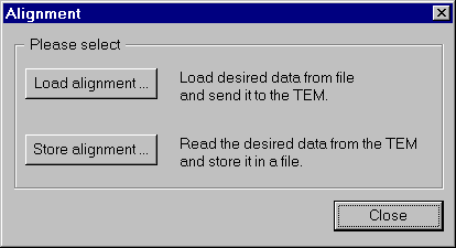
Clicking on opens the select alignment action dialog.
Select alignment action dialog
Clicking opens the 'Load Alignment' dialog. In this dialog you can select between Alignment data, Mode data and Stigmator data.
If stigmator data is selected, you can select one or more of the 3 stigmator coils (C2, Dif and Obj) to be reloaded. A click on LOAD opens the file selector dialog.
Here you have to specify a previously saved alignment file ( *.ali ), a mode file ( *.mod ) or a stigmator file ( *.stg ) respectively.
Mode data and stigmator data are valid immediately after reloading the data to the EM, whereas the alignment is not activated automatically. To activate
the alignment data settings the remote control has to perform a specified sequence of pressing and releasing buttons on the EM. Check the option
Activate alignment after reloading to execute that sequence after reloading the data.
Note: Loading/saving of alignment/mode/stigmator data is only available for Philips CMxx series microscopes.
Warning: Do not reload aligments of older CMxx software versions. You could misalign the EM drastically !
The files are not compatible between different versions !
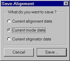
Clicking opens the 'Save Alignment' dialog. In this dialog you can select between Current alignment data,
Current mode data and Current stigmator data to be saved to file. After clicking OK a file selection dialog opens and you are requested to
enter a filename with suffix *.ali for alignment data files, *.mod for mode data files and *.stg for stigmator data files.
The 'Load Alignment' and 'Save Alignment' submenus
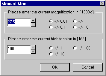
6.4.8 Manual Mag...
If the EMMENU is configured to use no remote control, the button
is enabled. A click on it opens the Manual Mag dialog.
'Manual Mag' dialog
This is the same dialog that also appears when EMMENU is started. You have to tell EMMENU every magnification or high tension change
you perform at the TEM via this dialog, otherwise strange effects ( like wrong scalebars, etc. ) will occur.
The radio buttons labeled +/- NNN define the increment or decrement, respectively, the spin controls will add to the
magnification or high tension value.
Note: The magnification value is in units of [1000x], i.e. a value of e.g. 27.5 represents a magnification of 27500.
The high tension's unit is kV. I.e. a value of e.g. 100 means 100kV.