
|


|
Giuseppe Destito
destito@scripps.edu
Pratik Singh, Ph.D
prasingh@scripps.edu
Kris Koudelka
koudelka@scripps.edu
Diane Thomas
dthomas@scripps.edu
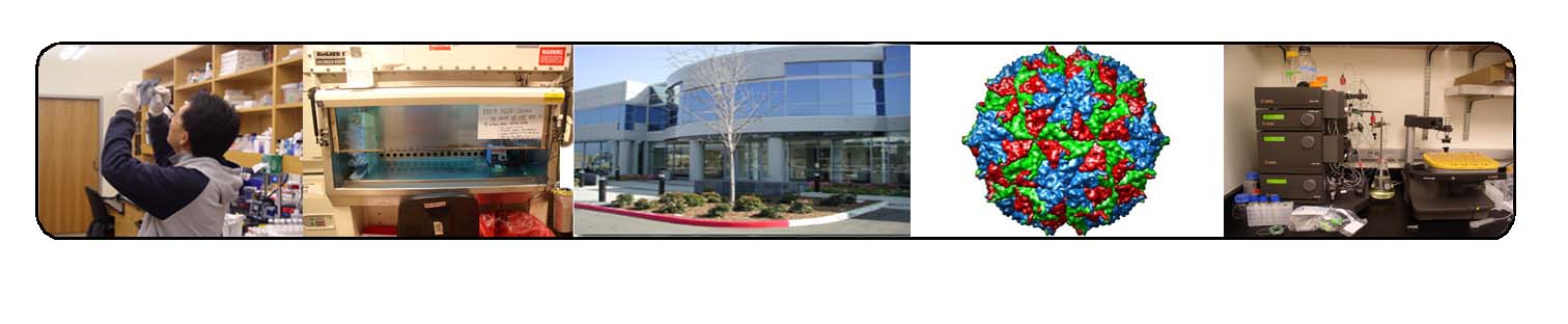
CPMV as a novel biomolecular sensor for vascular imaging
CPMV is an icosahedral, 31nm particle that is produced easily and inexpensively in black-eyed pea plants. In contrast to most other nanomaterials, the structure of the CPMV capsid is defined and can be engineered to display peptides or proteins in controlled orientations on particle surfaces via either genetic manipulation of the viral genome or by chemical attachment to the particle surface. CPMV is bioavailable and non-toxic, and the capsids are highly stable to temperature, pH, and chemical reaction conditions.
By conjugation to surface lysine residues, CPMV may be labeled with fluorophores at high densities, resulting in an extremely bright, non-toxic material that is an outstanding tool for imaging vasculature in live animals. We have shown that CPMV can effectively image the complete embryonic vasculature in several species and is superior to other imaging particles such as lectins, fluorescent dextrans or polystyrene microspheres. CPMV particles have proven highly useful for highlighting the process of angiogenesis in developing tumors. Uptake of particles into endothelial cells occurs, yielding a very bright imaging signal that can also differentiate between arterial and venous vessels. Such endothelial uptake is mediated by a cellular membrane protein, and uptake can be observed in the endothelium of a variety of healthy tissues as well as in disease states such as atherosclerosis.
Home Page · Schneemann Lab · Manchester Lab · Contact Us
Members Only · TSRI Home Page
|
|
|