


- VOL 17. ISSUE 36
November 27, 2017
SEARCH NEWS & VIEWS

New Painkillers Reduce Overdose Risk
High-Res Imaging Reveals How Cells ‘Take Out the Trash’
A Structural Clue to Attacking Malaria’s ‘Achilles Heel’
Potential New Autism Drug Shows Promise in Mice
Ozanimod Successful in Clinical Trials for Multiple Sclerosis
NEWS & VIEWS HOME
PAST ISSUES
KUDOS
SCIENTIFIC CALENDAR
CA AUDITORIUM EVENTS
CONTACT

- Palm Beach Post
Overdose-proof opioid? Scripps’ new drug could reduce deaths - Fierce Biotech
Identifying new ‘template’ for potent vaccine against HIV


FOLLOW US


High-Res Imaging Reveals How Cells ‘Take Out the Trash’
By Madeline McCurry-Schmidt
A new structural study shows exactly how cells take out the trash.
Using a high-resolution imaging technique called cryo-electron microscopy (cryo-EM), scientists at The Scripps Research Institute (TSRI) have revealed how cells degrade damaged or misfolded proteins in mitochondria—cellular compartments that serve as the “powerhouses” of the cell.
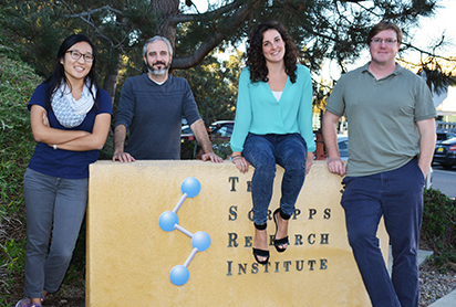
Left to right: Study authors Mia Shin, Gabriel Lander, Cristina Puchades and Luke Wiseman (Photo by Madeline McCurry-Schmidt)
Protein degradation is constantly occurring in cells, and is essential to all life forms because it allows cells to get rid of old proteins so that new ones can be made. Defects in protein degradation are closely linked to a variety of disorders, including cancer and Alzheimer’s disease.
Using cryo-EM, researchers solved the structure of YME1, a molecular machine that degrades proteins inside mitochondria. This structure allowed them to determine exactly how this machine uses molecular energy to chomp through dangerous proteins. “We’ve known for a long time that ATP molecules power protein degradation, but people never really understood what was going on at the molecular level.” said TSRI Graduate Student Cristina Puchades, first author of the study.
The new research was recently published in the journal Science.
Catching Mitochondria in the Act
Within cells, mitochondria are membrane-shrouded containers, so their interior is inaccessible to the protein quality control machinery used by the rest of the cell. Therefore, mitochondria have developed their own systems to manage protein levels.
“Mitochondria regulate their protein levels differently than the rest of the cell, and this structure substantially advances our understanding of this sophisticated process” said TSRI Associate Professor Gabriel Lander, Ph.D., who co-led the study with Steven E. Glynn, Ph.D., of Stony Brook University.
A graduate student in Glynn’s lab, Anthony Rampello, engineered the YME1 machine to enable this study. It then took the researchers at TSRI three years to solve the 3D structure of YME1 frozen in action, trapped in the process of degrading a protein.
“Our structure of YME1 bound to substrate reveals its mechanism of action and explains 30 years of biochemical research in ATP-driven unfoldases,” said TSRI Associate Professor Luke Wiseman, Ph.D., co-author of the study.
The YME1 machinery appears to chop up protein substrates in a two-step process. First, six ATPases surround the protein substrate slated for degradation, forming a structure around it that resembles a spiral staircase. The ATPases then thread the substrate through the center of the spiral staircase and feed it to a “degradation chamber” where the protein is cleaved into small bits.
The scientists were surprised to discover how this threading works. They found that the ATPase encircles the protein substrate, much like a spiraling staircase wrapping around a central pole. Each "step" of this staircase contains an amino acid that slots into the substrate protein in a zipper-like configuration. In this way, the staircase has a tight hold on the central substrate protein. The energy of ATP molecules fuel motions that cause the staircase to move like a “twisted escalator,” pulling the substrate downward.
As each escalator step reaches the bottom, its amino acid releases its grip on the protein substrate, and the step leap-frogs upwards back to the top of the staircase and takes a hold of the substrate for another round of downward motion. The authors compared the process to pulling a rope hand-over-hand.
Looking Toward Human Health
This study focused on the YME1 machine found in yeast, and the next step for the researchers is to solve the structure of a human version of YME1 that sits on the other side of the mitochondrial membrane.
In humans, mutations of this protein cause neurodegeneration in patients suffering from optic nerve atrophy and spastic paraplegia.
“Protein quality control and its impact on mitochondrial function are involved in many different sorts of neurodegenerative disorders,” said Puchades. “Now we can start using what we’ve learned in the context of disease.”
Additional authors of the study, “Structure of the mitochondrial inner membrane AAA+ protease YME1 gives insight into substrate processing,” are Mia Shin at TSRI and Christopher J. Giuliano of Stony Brook.
The study was supported by an American Heart Association predoctoral fellowship, the National Institutes of Health (grants DP2EB020402, S10OD021634, T32GM008468 and R01GM115898) and the Searle Scholar and Pew Scholar programs.
Send comments to: press[at]scripps.edu
