

New Service Offers Molecular Models a.k.a. "Thinking Tools"
By Mika Ono
BioMedical Graphics and the laboratory of Professor Art Olson have teamed up to offer the Scripps Research community a new molecular modeling service—not the kind of modeling that creates a virtual image on your computer screen, but the kind that builds an actual three-dimensional physical structure you can hold in your hand and pass from person to person.
The new service, which is being offered on a trial basis, grew out of the popularity of some of the models Olson was building as part of his molecular graphics research program on the La Jolla campus.
"People kept saying to me, 'I'd love to have a molecule like that,'" says Olson. "Now, there's a way they can get one."
The tangible 3-D models are most useful as "thinking tools," according to Olson, as well as for communication and teaching, for example in Scripps Research graduate classes.
"The feedback I get from researchers who have these models is that they're amazed by how much the models are used," says Olson, who has been beta testing the service for several months. "A physical object is persistent. It's there. In contrast to virtual representations, people contemplate physical objects longer and interact with each other around them more."
Olson also notes that a 3-D model engages more of one's senses than a virtual image, and thus more neurons in the brain. Studies in the perception and cognition literature support the idea that handling an object affects people differently than simply seeing it.
In fact, the history of molecular modeling goes back hundreds of years, and includes such pioneering figures as Linus Pauling and James Watson and Francis Crick. Over the last generation, though, as computers became commonplace and scientists' understanding of molecular structures became more complex, computer imaging gradually became the dominant tool for visualization.
"Now people are so used to seeing images on the computer that building a physical model can present challenges," Olson observes. "The prospect of making a solid model forces people to think about their molecule's physicality. They are confronted with such basic questions as 'Will it hold together?'"
While Olson's system builds on a long tradition, it also takes advantage of the latest in technology. Software is used to process atomic coordinates (generated by computer modeling, x-ray crystallography, or nuclear magnetic resonance) or volumetric images, such as from electron microscopy. This information is then used to build a geometric model that can be fed into a three-dimensional fabricating printer that "prints" solid objects out of thousands of layers of a special fine plaster powder.
As thousands of layers are built up, colored ink is spread in a pattern that is the cross-section of the physical structure on each plane. The ink acts as a binder, solidifying the part of each layer that will become the final object, and fusing it with the layer below. When the process is finished, the physical three-dimensional model is imbedded in the loose unbound powder, and can then be lifted out, dusted off, and finished with wax.
"This is advanced technology," says Olson. "No one will have it in individual labs any time soon, so I'm hopeful that people will find the new service useful."
Olson has trained Jon Huntoon, who works in his lab and studies biology and media at the University of California, San Diego, to use the system. Huntoon, who enjoys learning about the molecules scientists ask him to print, will staff the new service for Scripps Research investigators.
Bob Turner, director of BioMedical Graphics, adds, "We're excited to be working with Art Olson on this collaboration and we would like to thank him for donating the technology and the training to make this new service possible. As always, BioMedical Graphics is here to serve the Scripps Research community."
A lab's cost to create a model of a molecular object—which can range from small organic molecules to large biomolecular complexes, such as the ribosome, DNA polymerase, and virus structures—will depend on the model's size and the materials used, as well as the labor needed to produce a structure that can be built and handled. Researchers can minimize costs by generating a computer image using the Python Molecular Viewer (PMV) or any other software that outputs .stl, .ply, or .wrl files.
For more information about the new service, contact Jon Huntoon at huntoon@scripps.edu.
Send comments to: mikaono[at]scripps.edu
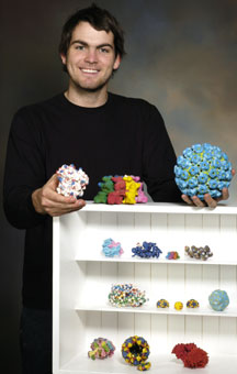
Jon Huntoon can produce solid molecular models for members of the Scripps Research community.
